

- Olympus cellSens Software

- Product Detail
- Company Profile
The Olympus cellSens platform creates a uniquely personal and intuitive imaging experience based on the operator’s preferred workflow. cellSens provides full control over the display and placement of icons, toolbars and controls to simplify the desktop and enhance productivity. cellSens is easy to use, powerful and flexible. Using a modular design, the software can be tailored to a budget as well as to the intended imaging applications. This enables the software to grow and adapt to meet evolving research needs.
The cellSens software package is not for clinical diagnostic use.
cellSens Packages
cellSens Entry
cellSens Entry is the ideal stepping stone for researchers wanting to move into digital image acquisition and documentation. By providing control over the camera via the unique customizable interface, Olympus cellSens Entry guarantees that each and every user can capture the images that they require using the settings that they want.
cellSens Standard
The Olympus cellSens Standard software builds upon the cellSens Entry package, taking acquisition beyond a single image, with advanced image capture processes (e.g. time lapse) and control of motorized and encoded microscope components. Measurement functions are more extensive, with many available morphological parameters, and freely editable measurement objects that don’t affect your original data. You can export your results to Microsoft Excel with just a single click. At any time cellSens Standard can be expanded with optional software Solutions which will enable further advanced functions. Panoramic imaging and extended depth of focus will free the microscope from its optical limitations while the NetCam function will allow easy and free projection of live images through any local network or Internet, for collaboration and education purposes.
cellSens Dimension
The most versatile member of the Olympus cellSens family is cellSens Dimension, featuring the unique and distinguishing Graphical Experiment Manage (GEM). Full control for complex research acquisitions and solid reliability for beginners can both be achieved by designing the experimental scheme with a few mouse clicks, with almost no need for training. Define rich experiments with automated loops, user interaction, and full hardware control and automatically create multiple results from a single experiment. cellSens Dimension further builds on the features available in cellSens Standard by including Z-stack acquisitions, auto-focus functions, and real-time deblurring of the live image. Together with the capability to control third-party high-end cameras and light sources, the acquired data is quickly processed with advanced analysis tools and then easily transferred into customizable reports, which are easily exported directly to Microsoft Word with the included Report Composer tool. Optional software Solutions are also available, implementing advanced and dedicated analysis functions such as FRET and FRAP.
cellSens Dimension Desktop
The Olympus cellSens Dimension Desktop software allows you to take your image processing offline, freeing up microscope acquisition systems to continue collecting data.
Image Acquisition and Experiment Design
Advanced Image Acquisition with Straightforward Operation
Graphical Experiment Manager (GEM)
Dimension
GEM is a flexible drag-and-drop interface to build simple or complex experiments within cellSens software. Combine actions within specialized frames to dictate the order and priority of automation. Easily acquire multichannel, z-stacks, and time-lapse images across one or more sample positions. GEM permits users to interact with the system during long timelapse imaging without terminating the experiment. Two-channel simultaneous imaging is also possible in GEM, allowing full utilization of all available hardware components.
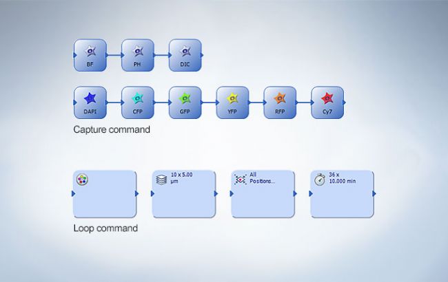
Capture Multidimensional Images
Dimension
Standard
The Process Manager makes it easy to capture multicolored and timelapse images with just a couple of clicks when imaging with a motorized stage. Z stack imaging is also possible when using a cellSens Dimension license.
Dimension + Multiposition
Standard + Multiposition
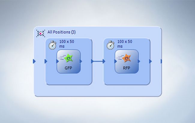
The optional multiposition solution is used to automatically capture multipoint and large area images.

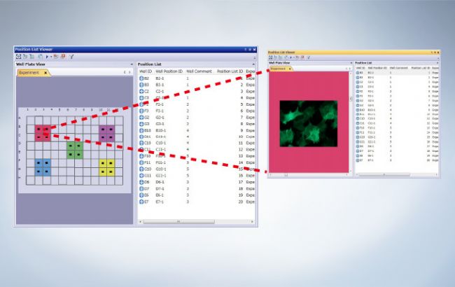
Well Plate Navigator
Dimension + Multiposition + Well Plate Navigator
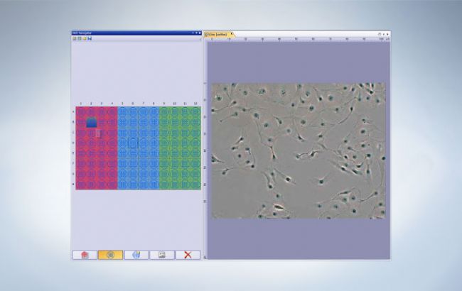
The Well Plate Navigator automatically scans and acquires images from standard and customized well plate formats. All acquired images, sample positions, and user comments can be saved into a structured database for rapid centralized access. Navigation to the center of any well is as simple as a single click. Wells can be selected individually, by row or column, or in discontinuous groups. Apply unique multidimensional acquisition settings to a single well or multiple selected wells in one step. The Well Plate Navigator can execute multiple experiments within a single well plate in support of complex experiments.
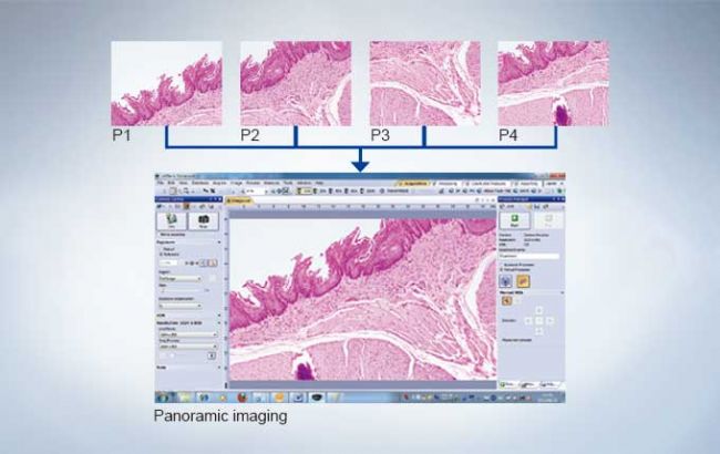
Real-time Panoramic Imaging
Dimension
Standard + Manual Process
Create stitched images in real-time with the Manual Process solution. Manual Process Control is available as an option for cellSens Standard software and included within cellSens Dimension software.
Motorized Panoramic Imaging
Dimension + Multiposition
Standard + Multiposition
With cellSens Dimension software, wide area imaging using a motorized stage is fully automated with the optional Multiposition solution. When combined with a motorized Z, this function can correct for the effects of sample distortion and tilting.
Extended Focus Imaging
Dimension
Standard + Manual Process
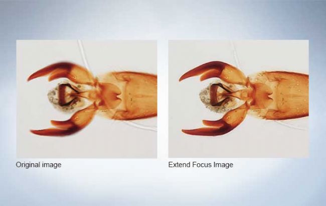
Create a single in-focus image from successive image planes as the focus knob is turned using the Extended Focus Imaging (EFI) function. A motorized focus drive fully automates EFI acquisition. EFI composite images can also be created directly from previously captured Z-stacks.
Sophisticated Image Processing
Spectral Unmixing
Dimension
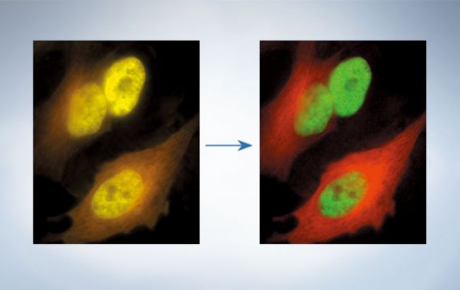
With the linear unmixing algorithm in cellSens Dimension, fluorochromes which overlap in their emission spectra - such as GFP and YFP - can be readily separated to produce crosstalk-free fluorescent images. This linear unmixing tool can also separate autofluorescence-related background signal.
Deconvolution
Dimension
cellSens Dimension includes Live 2D deblurring for image preview and acquisition, to enable better focusing on thick specimens.
Dimension + Cl Deconvolution
The optional CI Deconvolution Solution employs the latest Constrained Iterative Deconvolution algorithms to produce improved resolution, contrast, and dynamic range with industry-leading speed. by using GPU processing.
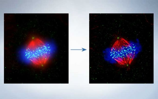
Cell line: Human cervical cancer cell line (HeLa)
Immunostaining: Hec1 staining (green, Alexa Fluor-488), a-tubulin staining (red, Alexa Fluor-568), DAPI staining (blue)
Mitotic HeLa cell derived from human cervical cancer. Mitotic spindle and kinetochores are stained with anti-a-tubulin (red) and anti-Hec1 (green) antibodies, respectively. Chromosomes interact with microtubules constituting mitotic spindle via kinetochores, protein structure assembled on centromere region of chromosomes.
Image data courtesy of: Department of Molecular Oncology, Institute of Development, Aging, and Cancer, Tohoku University Masanori, Ikeda and Kozo Tanaka
Best Focus Extraction
Dimension
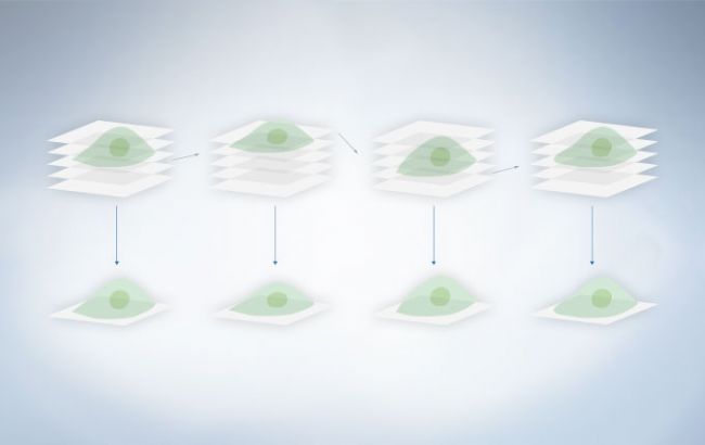
Extract the best focus from images, including z-stack & time-lapse images. This function is effective in creating T-series images with the best focus possible, even when working with defocused time-lapse images.
High Dynamic Range (HDR) Imaging
Dimension
By automatically capturing many images at different exposures the HDR function creates a final image with a much greater dynamic range, where low intensity signals are clearly visible without overexposing the bright areas of the sample.
Camera and Device Solutions
High End Camera Solution for cellSens Dimension
Dimension
Following the needs of advanced researchers cellSens Dimension can control of some of the latest sCMOS and EM-CCD cameras. Additional controls are added to the Camera Control Panel to allow effortless usage of these advanced cameras features in the usual cellSens interface.
High End Device Solution for cellSens Dimension
Dimension
cellSens Dimension utilizes advanced devices, like image-splitters and piezometric motorized Z axis devices, obtaining a substantial increase in the maximum acquisition speed. Together with the control of the Yokogawa CSU-W1 spinning disk confocal unit this solution allows cellSens users to follow fast and dynamic event in their live samples, obtaining reliable results together with the Olympus Real Time controller.
Image Analysis and Tools
Versatile Analysis Methods and Macro Tool
Manual Object Counting and Classification - Touch Count
Dimension
Standard
The cellSens touch count feature allows convenient manual cell counting. Cells or other objects are marked upon a mouse click, counting and automatically assigned to a user defined object class.
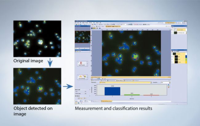
Automatic Object Detection for Measurement and Classification
Dimension
Efficient and precise threshold-based object detection for automated
nuclei counting, as well as classification are possible. Results can be conveniently exported to Microsoft Excel for further analysis.
Dimension + Count & Measure
Standard + Count & Measure
The extensive set of manual measurements in cellSens can be further expanded with the Count & Measure Solution. Easily perform automatic object measurement and classification in an interactive interface where recognized objects are always linked with their measurements.

Macro Manager
Dimension
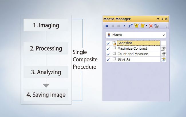
Use the macro manager to automate typical acquisition and data analysis workflows. Macro commands can be applied to multiple images simultaneously and can reduce the time required to complete complex imaging and image analysis.
Image Comparison (simultaneous image windows)
Dimension
Standard
Display images side-by-side for accurate comparison, with simultaneous zooming and movement.
Manual Measurement
Dimension
Standard
Distances between points, areas, intensity measurements, and morphological parameters are accessible using the cellSens software measurement tools. Measurement data are saved as an image layer that can be exported to MS Excel and cellSens workbook formats, or viewed using OlyVIA, a free image viewer software package.
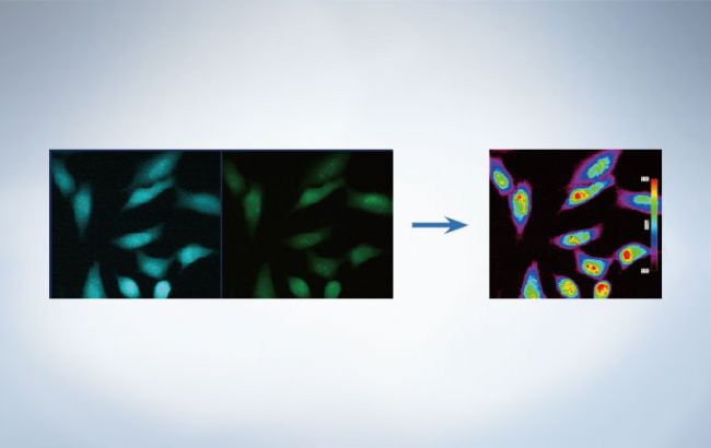
Intensity Analysis
Dimension
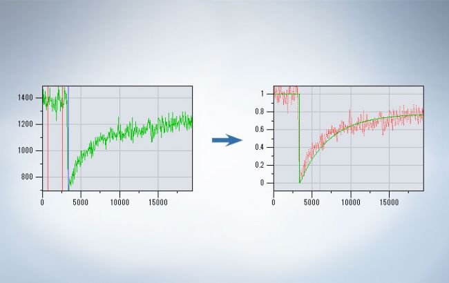
Graphically depict intensity and ratio values defined by Regions of Interest (ROIs) and adjust ROI placement to compensate for cell movement. Convert variations of intensity to hue and brightness using Intensity Modulated Display (IMD) to visually enhance the fine image structures often found within ratio or FRET images. The Ratio/FRET Solution is used to display and analyze real-time ratiometric imaging and data. FRET analysis of both sensitized emission and acceptor photo-bleaching is also supported within this user friendly workflow. The Photo-Manipulation Solution can be used for the curve-fitting analysis of FRAP images.
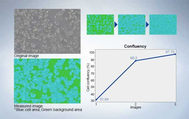
Confluency Checker
Dimension
Standard + Confluency Checker
Olympus’ cell identification algorithm enables measurement of cell count and confluency on phase contrast images. It is possible to measure multiple images at once, thus data averaging and total cell count estimation are easily measured across an entire vessel. A cell growth curve can be output by measuring along with time series.
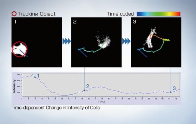
Object tracking
Dimension + Count & Measure + Tracking
The cellSens Tracking Solution provides and intuitive tool to detect, track, and analyze moving objects, including dynamic processes such as cell movement and division.
Bioluminescence of RA-induced differentiating cells at day 12 from Bmal1:luc stably transfected ES cells
Image data courtesy of: Kazuhiro Yagita, M.D. Ph.D. Department of Physiology and Systems Bioscience, Kyoto Prefectural University of Medicine
Reference: Proc Natl Acad Sci U S A. 107(8): 3846–3851(2010)
Reporting and Database
Reporting
Dimension
A convenient Reporting tool combines images with metadata measurement data, and user-customized fields into a report template with easy drag-and-drop operation. These Microsoft Word* reports provide the ability to quickly and easily collaborate with colleagues and communicate results.
*Requires Microsoft Word version 2010 or later
Database
Dimension + Database Core or Database Client
Standard + Database Core or Database Client
Entry + Database Client
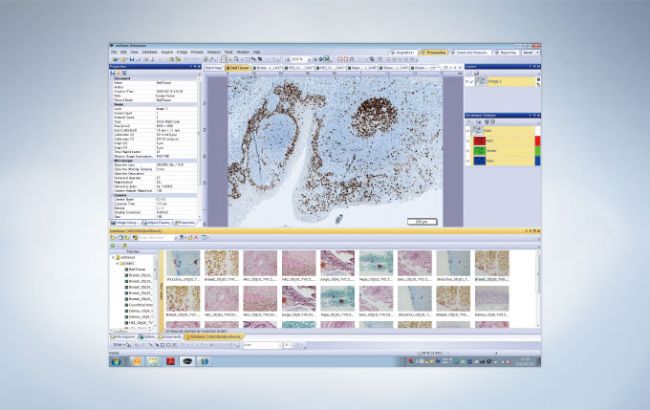
The Database Core Solution allows the creation of user-defined databases, with full access control, which can be shared across a network. The database not only collects images but also all associated image properties, user comments and any kind of related files, like spreadsheets for other documents. An interactive query tool makes it easy to find the desired data, with automatic preview of the found images. With the Database Client Solution it is possible to conveniently read and write to the shared database from many different stations.
Database + Well Navigator
Dimension + Multiposition or Well Plate Navigator
Database Core or Database Client
Standard + Database Core or Database Client
+
Entry + Database Client
In combination with the Well Navigator Solution, the Database Solution greatly improves the efficiency of viewing and analysis of well plate images with a large amount of data. By clicking on icons for image information such as the date, file name, or well plate number, any selection of captured images can be viewed for further analysis. Together, they allow viewing of captured images and continuous analysis of selected images (the Batch Macro function) via the well plate interface.
Expandability
Remote Live Image
Dimension + NetCam
Standard + NetCam
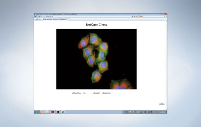
Using standard TCP/IP protocols, the cellSens NetCam Solution facilitates the transfer of live, as well as stored, images throughout a network for teaching, mentoring or supervision. As a result, even when outside of the laboratory, colleagues and/or supervisors can monitor the work from any point on the network, improving the efficiency of the laboratory
Click here for the details of NetCam
Solution
Each cellSens Package can be expanded towards a specific application by using optional “Solutions”
Dimension
Multiposition Count & Measure CI Deconvolution Well Plate Navigator Ratio / FRET
Photo Manipulation Database Core Database Client NetCam Life Sciense Analysis
Standard
Multiposition Count & Measure Manual Process Database Core Database Client
Confluency Checker NetCam
Entry
Manual Process Encoded Device Database Client Interactive Measurement
*cellSens Entry is not available in all areas.
Personalization and User Interface
Clinical Research
Simple Layout
Entry
Standard
Dimension
Novice and expert users will simultaneously benefit from the "Simple Layout," an interface designed for a clinical research workflow. Acquire, annotate, share, and save your images using the intuitive Smooth Control tool window. Built-in measurement tools display only when required, reducing software clutter and minimizing distraction.
Conference mode
Entry
Standard
Dimension
Use Conference Mode to fill the screen with live or static images for presentation and collaboration with just one click. Graphic annotation tools are available at your fingertips for image markup without the need to exit Conference Mode, improving workflow efficiency and saving time.
Scientific Research
Dark Application Skin
Standard
Dimension
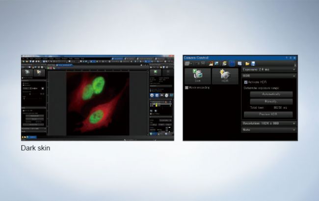
The Dark Application Skin reduces computer monitor-generated ambient light and allows cellSens users to adapt to darkened environments; icon contrast remains high for easy recognition and quick selection.
Customizable Window Arrangement
Standard
Dimension
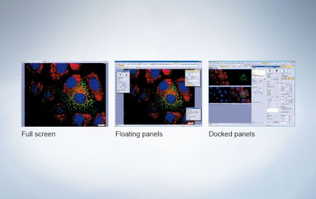
Tools and windows can be organized to suit the job at hand, optimizing the functionality of the layout.
Workflow Oriented Layout Tabs
Standard
Dimension
All common functions are integrated into tabs, combining all related options and settings. These layout tabs allow easy selection of functions according to the particular workflow. For example, camera control features are displayed under the Acquisition tab. These are then removed from view when switching to the Processing tab in the next stage.
My Functions
Standard
Dimension

cellSens lets users create and save custom toolbars for the most frequently used functions and then save them to the My Functions window.
-
Olympus is one of the world’s leading manufacturers of professional opto-digital products for medicine, science and industry. As a result, Olympus provides a comprehensive range of solutions. From microscopes for training and routine tasks to high-end system solutions in the fields of life science, there is a system for every need. The product line is complemented by innovative laboratory equipment for cellular research applications and the new all-in-one microscopes that offer user engagement at all levels.
| Request Information |
| Related Products |
| Recently viewed products |