

- Cell Analysis Software

- Product Detail
- Company Profile
NoviSight 3D cell analysis software provides statistical data for spheroids and other 3D objects in microplate-based experiments.
Not for clinical diagnostic use.
Some photos provided courtesy of Lawrence J. Ellison Institute for Transformative Medicine of USC
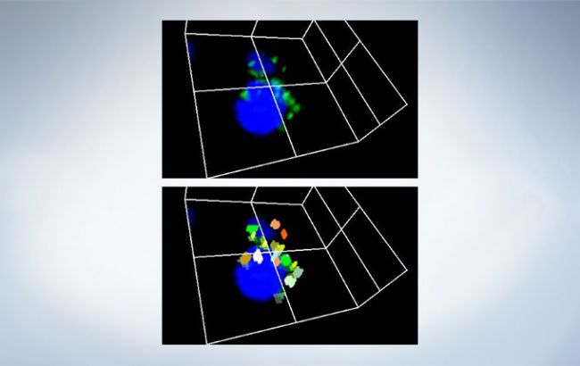
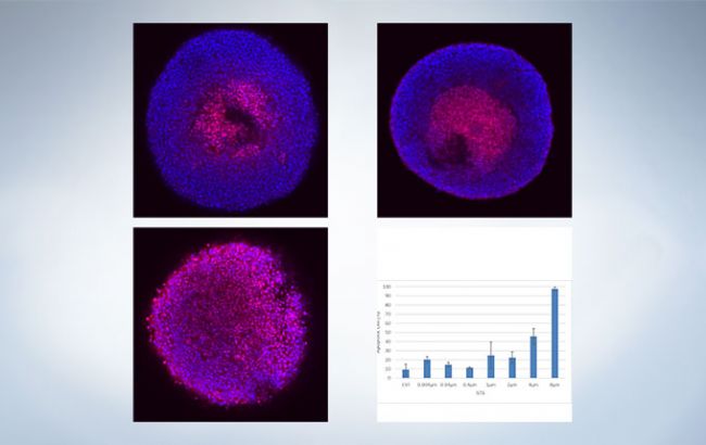
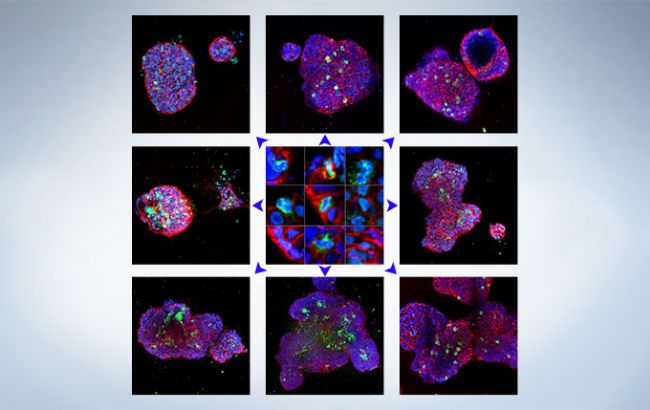
Accurate Result of 3D Analysis
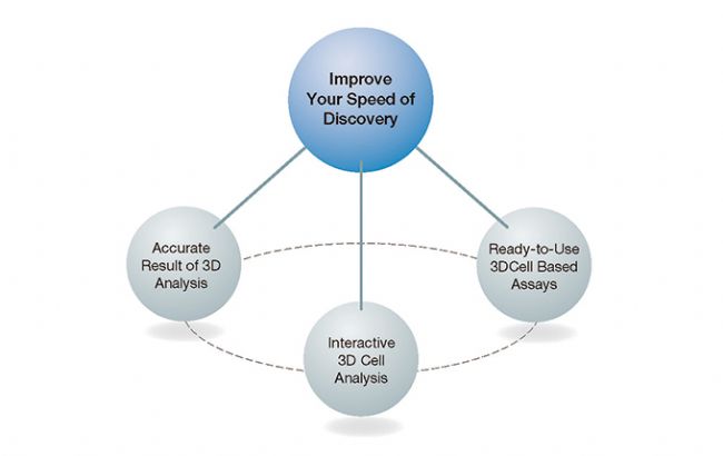
Designed for microplate-based experiments, NoviSight™ software’s True 3D technology enables you to check the morphology of your samples. Measure a range of spheroid or cell nuclei parameters including volume and sphericity. For biologists, the software makes it possible to measure and analyze physiological relevant 3D models, helping improve the speed of drug discovery.
See What’s Hidden in the Depth of 3D
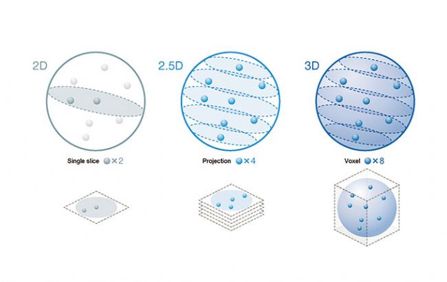
If you’re still using two-dimensional methods to analyze three-dimensional samples, you’re not seeing the whole picture.
NoviSight software enables you to more easily:
・Capture rare cell events
・Get accurate cell counts
・Improve the sensitivity of detection
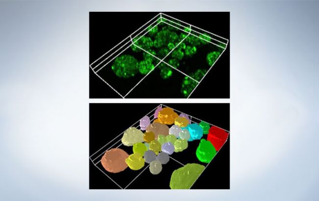
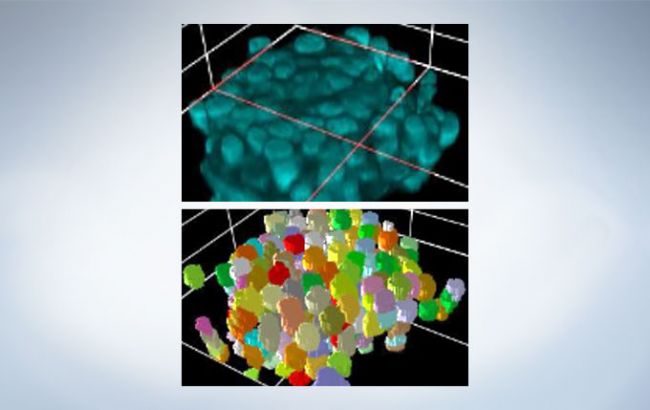
Fast, Precise Object Detection
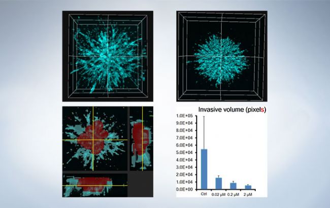
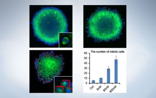
The software’s algorithms are designed to quantify cell activity and interactions in three dimensions. This precise object detection enables the software to analyze objects of interest to provide morphology and spatiotemporal parameters in 3D space.
Detect objects from whole structures to subcellular features and evaluate:
・Changes in spheroids
・Cell cytoplasm
・Nuclei
Imaging with Clearing Techniques
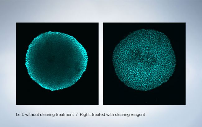
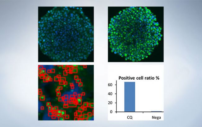
As shown in the example, treating with a clearing reagent such as Scale-S enables clear imaging deep inside 3D cultured spheroids.
Flexibility to Work with Different Imaging Techniques
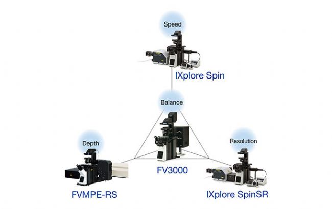
The software is compatible with all Olympus confocal imaging systems. No matter which system you use, the software will quantify and analyze the objects you capture.
Spinning disk: Sensitive and fast imaging for live cells, tissues, and small organisms.
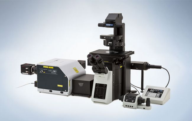
Super resolution: Fast confocal image acquisition with a large field of view, which is designed for experiments involving live cells with reducing phototoxicity and bleaching.
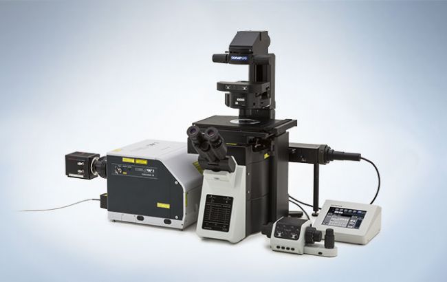
Live cell: Super resolution imaging device for live cell imaging with 120-nano meter resolution, observes the fine details and workings of internal cellular structures.
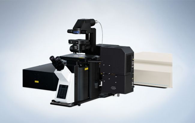
Deep imaging and cleared tissue(including cleared spheroids): Deep imaging in biological tissue reveals both detail and dynamics deep within the specimen.
Ready-to-Use 3D Cell-Based Assays
Reduce Your Assay Development Time
The software comes with a variety of ready-to-use 3D cell assays. Simply choose the assay that suits your experiment and begin analyzing. You can also automate sequential processes for multiple data sets and assays.
Easily Design Assays for Advanced 3D Cell Analysis
If a suitable assay isn’t readily available, you can design your own. Easily create assays for multi-well, multichannel, and time-lapse experiments.
Assay Fitting Service
For advanced 3D cell analysis, we can work with you to customize an assay for your experiment. The software’s algorithms can be easily changed so we can adjust the algorithm for you promptly.
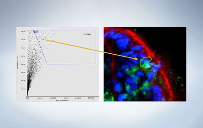
Reduce Your Analysis Time with Interactive 3D Cell Analysis
All the data you need—recognition, analysis, and statistics—are conveniently located in one intuitive user interface. The original images and quantitative data are linked for easy validation and interpretation. You can also export that data as a CSV or FCS file for further analysis.
All on One Screen
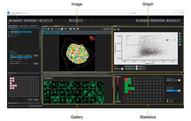
・Image: Get 2D or 3D views of your samples; you can locate objects in 2D within an image plane or switch to 3D to explore an entire spheroid
・Graph: The scatter plot makes it simple to classify objects; clicking and selecting an individual point brings up an image of that object
・Gallery: Observe the details in each region of an object at a glance; visualize how segmentation is working by highlighting two areas in the scatter plot and display the resulting galleries side by side
・Statistics: View quantitative results numerically or visually on a heat map
Data and Images Are Always Connected
NoviSight™ software’s accurate cell detection enables you to plot objects on a scatter plot or histogram. All the data are interactive—display your results in an image gallery, heat map, or table. Clicking a point in any of these display options shows the sample’s corresponding image.

-
Olympus is one of the world’s leading manufacturers of professional opto-digital products for medicine, science and industry. As a result, Olympus provides a comprehensive range of solutions. From microscopes for training and routine tasks to high-end system solutions in the fields of life science, there is a system for every need. The product line is complemented by innovative laboratory equipment for cellular research applications and the new all-in-one microscopes that offer user engagement at all levels.
| Request Information |
| Related Products |
| Recently viewed products |