

- Inverted Microscope

- Product Detail
- Company Profile
ENVIRONMENTAL CONTROL FOR LIVE SAMPLES
Maintain Cell Health Over Several Days
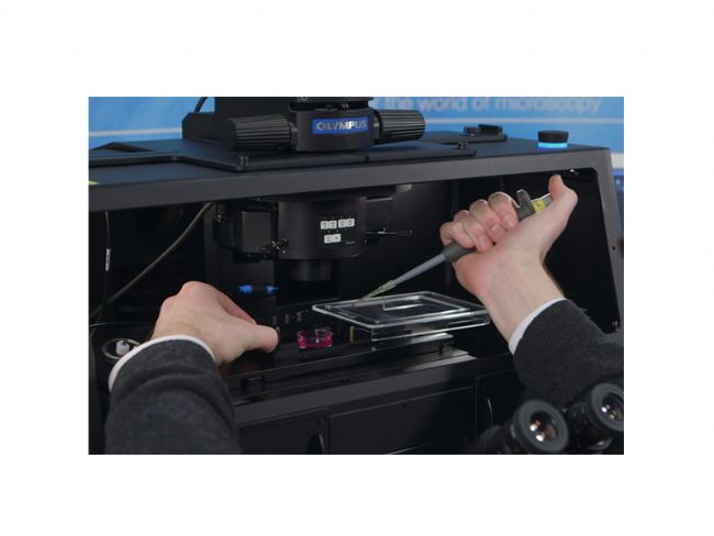
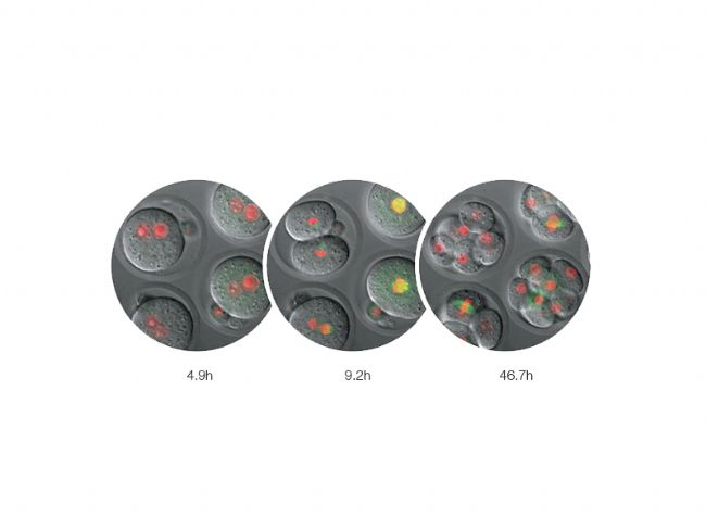
Box-type incubator* enables time-lapse observations over a period of several days, while the microscope CO2 incubator* can be fitted to the stage for two-day time-lapse observations, maintaining cell activity to significantly improve the reliability of time-lapse observation.
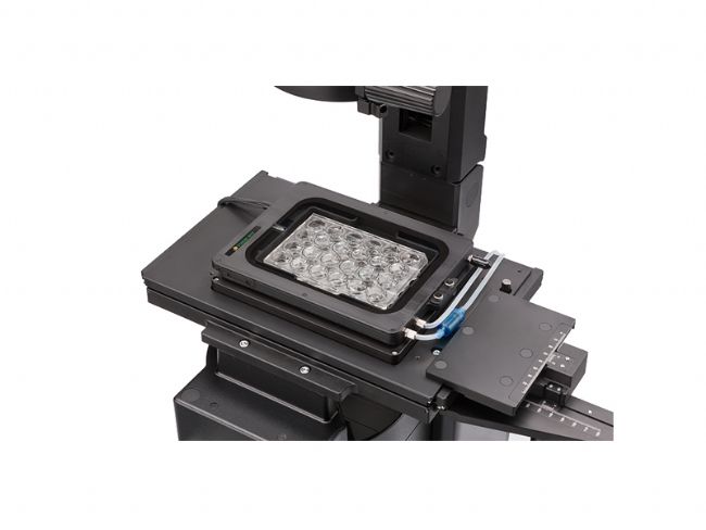
CO2 Stage Top Incubator*
Precise controls maintain a constant environment, with the dish or well plate controlling temperature, humidity, and CO2 concentration.
Incubator*
A box-type incubator* keeps the microscope temperature stable while safely enclosing many components.
*Third-party products.
HIGHLY-STABLE ENVIRONMENTAL CONTROL
In this SelectScience Interview, Jutta Bulkescher, Microscopy Specialist at the Center for Protein Research/Danish Stem Cell Center, University of Copenhagen, describes the wide range of research conducted at her facility and explains how the Olympus cellVivo incubation system is enabling her to reliably perform stem cell analysis, whilst maintaining cells under strict physiological conditions.
IMAGING STABILITY
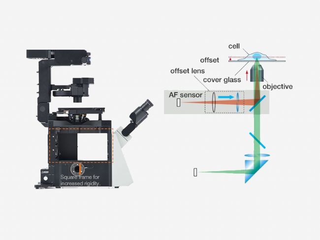
The frame architecture and focus drive design of the IX83 system offer enhanced rigidity that reduces the impact of vibration and temperature. It maintains desired positions along the X, Y, and Z axes to facilitate reliable time-lapse and multipoint imaging. When combined with the Olympus ultrasonic stage (IX3-SSU) and Z-drift compensator (IX3-ZDC2), the IX83 microscope can capture high-precision, multipoint time-lapse images that are aligned and in focus.
LIVE CELL IMAGING
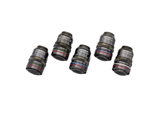
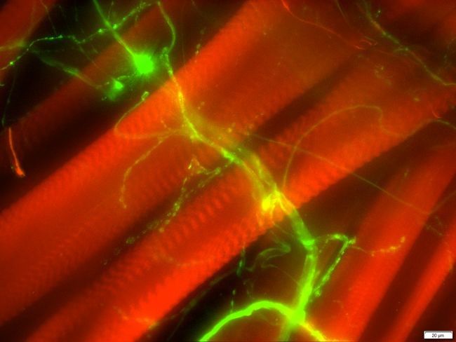
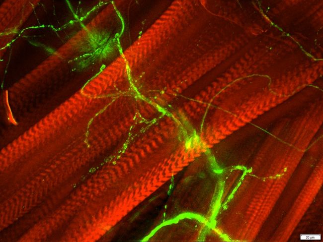
The optional Olympus Silicone oil immersion objectives enable optimal imaging of living cells over time. The refractive index of silicone oil (ne≈1.40) is close to that of living tissue (ne≈1.38), enabling high-resolution observation deep inside living tissue with reduced spherical aberration caused by refractive index mismatch. Silicone oil does not dry out or harden, so there is no need to add more oil, making it ideal for extended time-lapse observations.
FAST, MICROSECOND-ACCURATE DEVICES
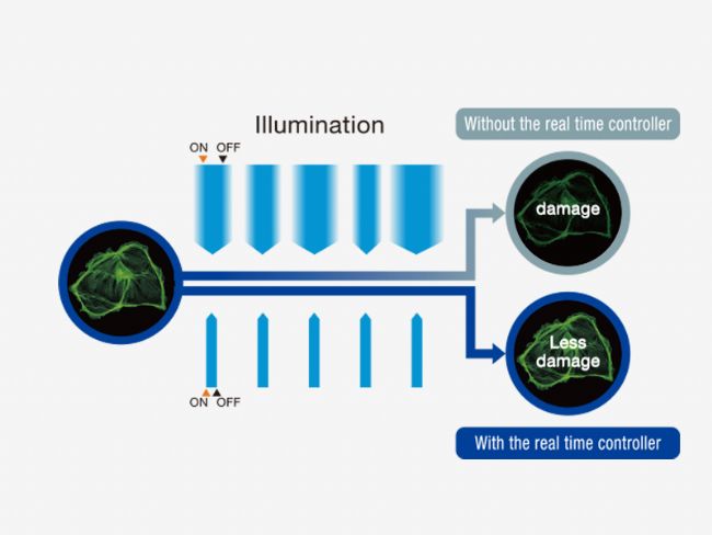
The fast filter wheel, shutter, LED light source control, and real-time controllers (U-RTC) enable less photobleaching and phototoxicity, therefore resulting in healthier cells and more robust data.
TRACKING
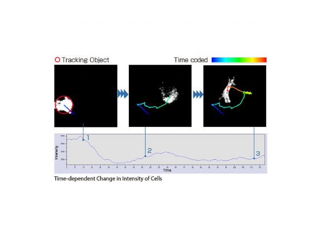
Precise tracking experiments are possible with cellSens tracking and count & measure solutions.
RAPID DECONVOLUTION
Olympus cellSens Dimension includes live 2D deblurring for live preview and acquisition, to enable better focusing on thick specimens. In addition, more advanced 3D deconvolution techniques are available to reassign out-of-focus light. The optional CI deconvolution solution employs a constrained iterative deconvolution algorithm to produce improved resolution, contrast, and dynamic range with industry-leading high speed by using GPU processing.
LARGE FIELD OF VIEW
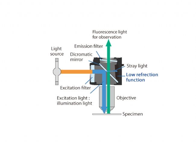
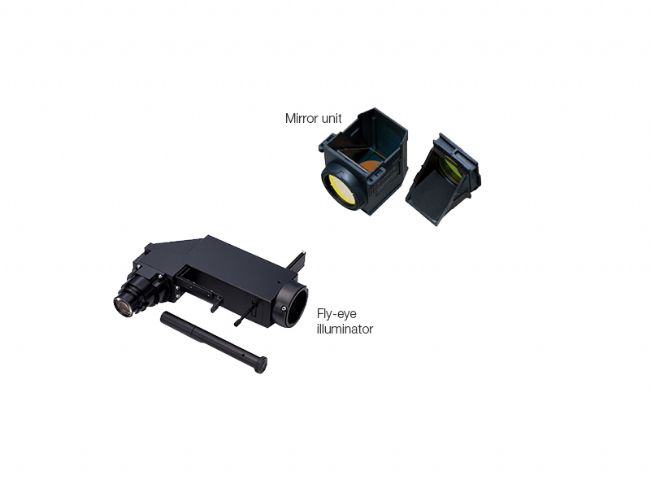
The large field of view Olympus optics, including mirror units and fly-eye lens systems, provide uniform fluorescence images and enable the use of sCMOS cameras with large sensors.
EASE OF USE
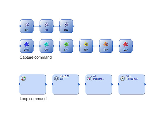
Fully automated multi-dimensional observation (X, Y, Z, T, wavelength, positions) with easy experiment set up using the Graphical Experimental Manager (GEM) in cellSens Dimension imaging software.
-
Olympus is one of the world’s leading manufacturers of professional opto-digital products for medicine, science and industry. As a result, Olympus provides a comprehensive range of solutions. From microscopes for training and routine tasks to high-end system solutions in the fields of life science, there is a system for every need. The product line is complemented by innovative laboratory equipment for cellular research applications and the new all-in-one microscopes that offer user engagement at all levels.
| Request Information |
| Related News |
| Other Products |