

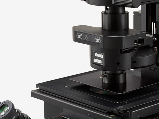
- Inverted Microscope

- Product Detail
- Company Profile
HIGH-SPEED CONFOCAL IMAGING
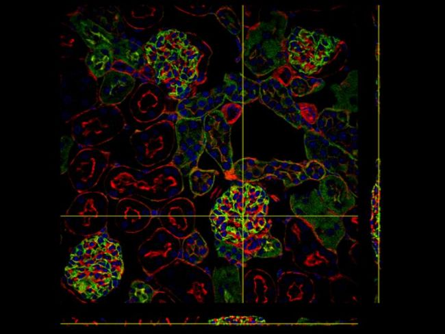
The spinning disk unit (YOKOGAWA CSU-W1) provides high-speed confocal image acquisition with a large field of view. Olympus cellSens software’s 3D deconvolution technology improves image resolution, contrast, and dynamic range, for striking high-speed 3D imaging.
*Image: Kidney Section Slide (Blue: DAPI, Green: WGA, Red: Phalloidin)
REDUCED PHOTOXICITY
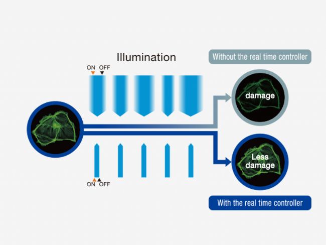
Designed for experiments involving live cells, the spinning disk (YOKOGAWA CSU-W1) reduces phototoxicity and bleaching. The Olympus real-time controller (U-RTCE) provides optimized device speed and precision during automated acquisition, while the Olympus Z-drift compensator (IX3-ZDC2) maintains focus at every frame, and reduces stress to the sample during data acquisition.
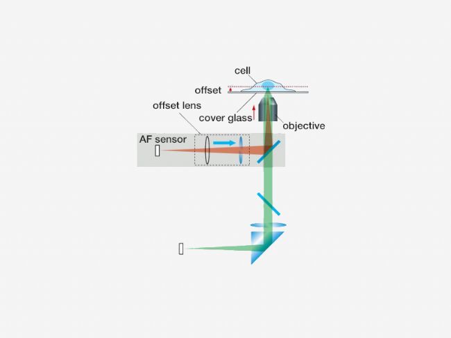
PRECISE 3D IMAGING
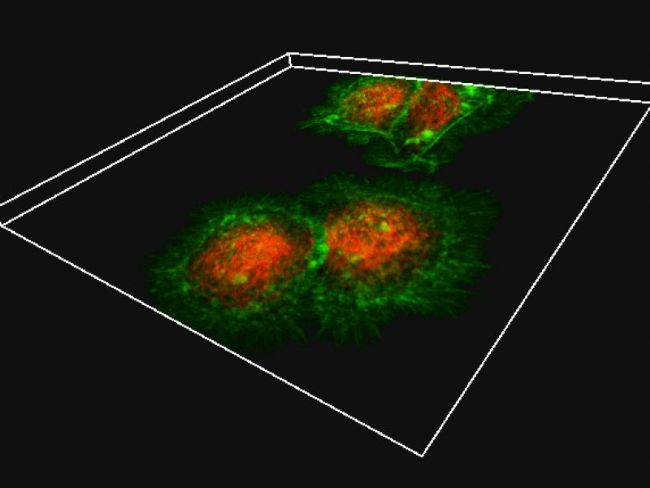
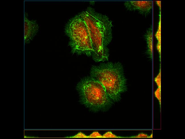
The pinhole geometry of the Yokogawa CSU-W1 spinning disk produces excellent image contrast at greater depth for deep imaging. In addition, the IXplore Spin system combines high NA silicone oil objectives with ergonomic spherical aberration correction collar adjustment for excellent light gathering and dimensional fidelity. These elements make the IXplore Spin system the right choice for researchers who need to image living cells at high resolution without sacrificing speed, accuracy, or image quality.
*Image: HeLa cells (Green: Actin, Red: Tubulin)
UPGRADE TO THE SPINSR SYSTEM
The IXplore microscope system is designed to meet your evolving research needs. The SpinSR super resolution module is available as an upgrade to any existing Olympus IX83 microscope and can be configured to fit your budget.
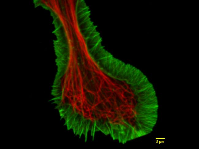
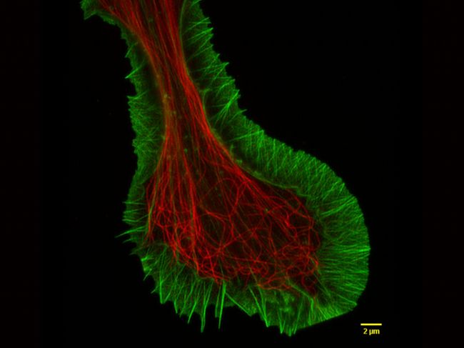
*Image: Fluorescent staining of microtubules (red: Alexa 594) and actin (green: Alexa 488 phalloidin) in growth cone of NG108 cells. Image courtesy of: Dr.Kaoru Katoh , Biomedical Research Institute, National Institute of Advanced Industrial Sciences and Technology (AIST)
SIMULTANEOUS MULTI-COLOR CONFOCAL IMAGING
The IXplore Spin laser combiner is scalable from two to six laser lines. A dual camera option is available to support simultaneous multi-channel imaging when higher speed and information bandwidth are required. Available excitation wavelengths include 405 nm, 445 nm, 488 nm, 514 nm, 561 nm, and 640 nm.
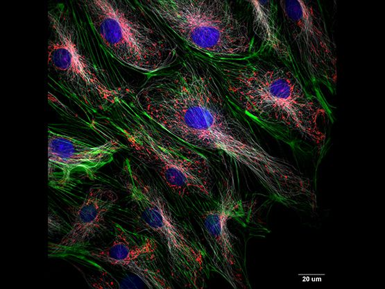
*Image: Rat-kangaroo Epithelial Kidney cells (Blue: Nuclear, Green: Actin, Red: Mitochondria, White: Tubulin)
HIGH CONTRAST UNDER BRIGHT CONDITIONS
The stage-top umbra unit is designed to eliminate the effect of stray light on fluorescence imaging and observation. The unit blocks room light to enhance the contrast of fluorescence and provide clear fluorescence observation under bright conditions.
-
Olympus is one of the world’s leading manufacturers of professional opto-digital products for medicine, science and industry. As a result, Olympus provides a comprehensive range of solutions. From microscopes for training and routine tasks to high-end system solutions in the fields of life science, there is a system for every need. The product line is complemented by innovative laboratory equipment for cellular research applications and the new all-in-one microscopes that offer user engagement at all levels.
| Request Information |
| Related News |
| Other Products |