

- Inverted Microscope

- Product Detail
- Company Profile
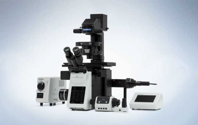
The fully motorized IX83 is the most advanced member of the IX3 series of inverted imaging systems and offers a new level of application flexibility. Choose from the one-deck system with a low, ergonomic stage or the two-deck system with additional expansion capabilities. Both provide the ability to perform a multitude of imaging applications, from long-term time-lapse imaging and other demanding techniques to routine testing and documentation. Users can mold the components and controls to best suit their workflow.
Expandable to Meet Growing Research Needs
The fully-motorized IX83 is designed to satisfy a variety of research needs. With additional modules providing expanded functionality, both microscope options enable a multitude of imaging techniques, ranging from casual documentation to long-term time-lapse imaging and other demanding techniques.
The unique open frame of the IX83 provides ready access to the light path, thus making it easy to add or change modules. A variety of deck modules can be easily exchanged to add or remove functions as needed. With a simple slide-in design, the IX3-ZDC2 Z drift compensator module can be easily added to any IX83 system to maintain continuous focus throughout an extended time-lapse experiment.
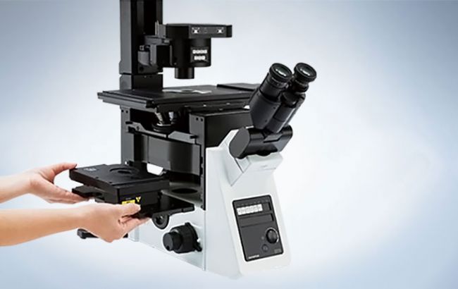
IX83: Two-deck System

Enables high-speed, fully automated device selection during live cell research and advanced image acquisition. The two-deck configuration provides excellent expandability.
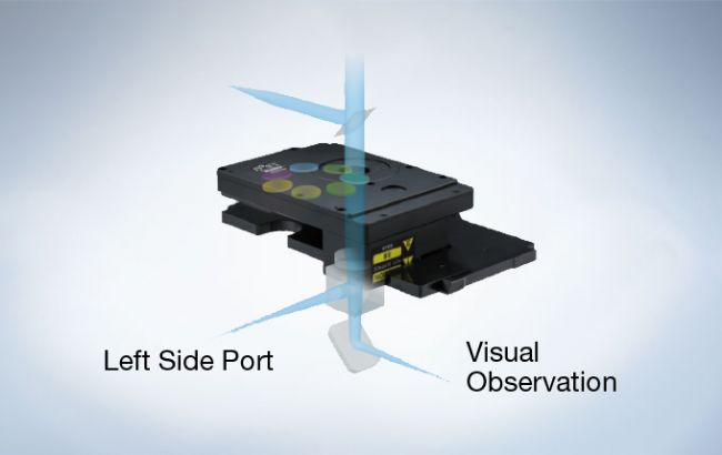
IX83: One-deck System
An intelligent, motorized microscope featuring a large field number (FN22, left side port) and IX3-ZDC2 compatibility, thus creating a new standard for live cell imaging.
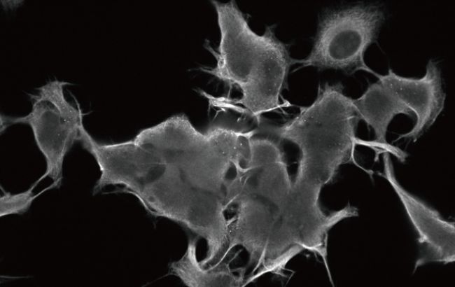
Reliable, Clear and Bright High-Resolution Images
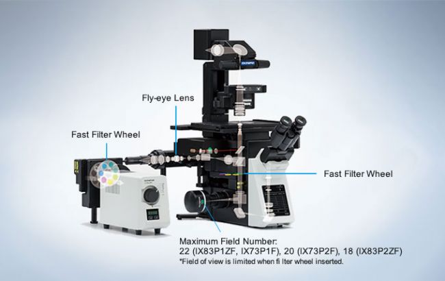
Olympus UIS2 infinity-corrected optics ensure high optical transmittance with a broad range of objectives. UIS2 optics feature wide chromatic correction and enable high resolution, high S/N primary images regardless of the observation method. The wide field of view and Fly-Eye lens system provide uniform fluorescence images and enable the use of sCMOS cameras with large sensors.
Excellent Image Quality
Apochromatic Objectives Enable High-Resolution Phase Contrast and Fluorescence Observation
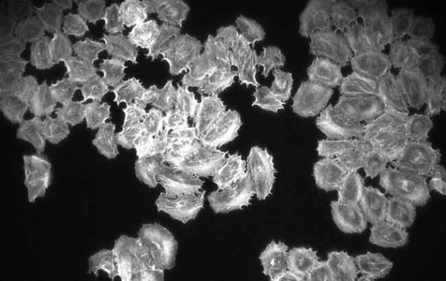
Phase contrast apochromatic objectives (UPLSAPO100XOPH, PLAPON60XOPH) enable high-precision imaging free from image shift, even during simultaneous phase contrast and fluorescence observation. This eliminates the need to change objectives when switching observation methods.

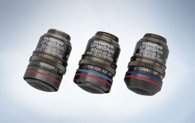
Silicone Objectives* Enable High-Resolution Observation Deep into Live Cells
Olympus offers three high-NA silicone immersion objectives:UPLSAPO30XS, UPLSAPO40XS, and UPLSAPO60XS. The refractive index of silicone oil (Refractive index: ne≈1.40) is close to that of living tissue (Refractive index: ne≈1.38), enabling high-resolution observation deep inside living tissue with minimal spherical aberration caused by refractive index mismatch. Silicone oil does not dry out or harden, so there is never a need to readminister the oil, making it ideal for extended time-lapse observations.
*Use dedicated silicone oil.

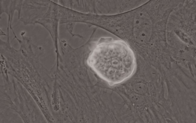
Special Objective Available for iPS/ES and Floating Cell Observation
This high-NA phase contrast objective (UCPLFLN20XPH) is especially suited for observation using plastic dishes. It enables high-resolution observation of the cell proliferation process and delivers improved contrast across a wide area.
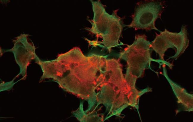
Bright, Uniform Fluorescence Illumination
The fluorescence illuminator (IX3-RFALFE) incorporates a Fly-Eye lens system to provide even light distribution. This provides bright and even illumination to the entire field, including the periphery of the visual field.
High S/N Fluorescence Mirror Units for Efficient Signal Detection
All fluorescence mirror units feature filters treated with a specially-developed coating that minimizes noise by absorbing more than 99% of stray light. The outstanding performance and high transmittance of the mirror units ensure efficient fluorescence signal detection.

cellSens Deconvolution
cellSens Dimension includes Live 2D deblurring for image preview and acquisition, to enable better focusing on thick specimens. Additionally, more advanced 3D deconvolution techniques are available in cellSens to reassign out-of-focus light. The optional CI Deconvolution Solution employs the latest Constrained Iterative Deconvolution algorithms to produce improved resolution, contrast, and dynamic range with industry-leading speed by using GPU processing.
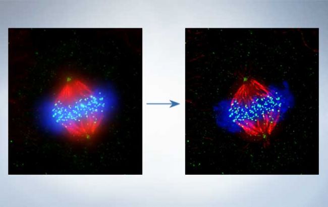
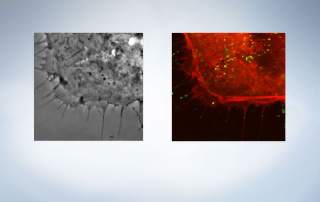
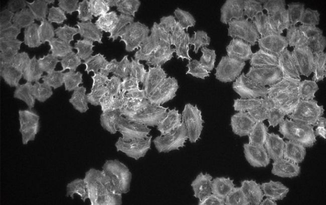
Cell line: Human cervical cancer cell line HeLa cell Immunostaining: Hec1 staining (green, Alexa Fluor-488), a-tubulin staining (red, Alexa Fluor-568), DAPI staining (blue) Mitotic HeLa cell derived from human cervical cancer. Mitotic spindle and kinetochores are stained with anti-a-tubulin (red) and anti-Hec1 (green) antibodies, respectively. Chromosomes interact with microtubules constituting mitotic spindle via kinetochores, protein structure assembled on centromere region of chromosomes.
Image data courtesy of: Department of Molecular Oncology, Institute of Development, Aging, and Cancer, Tohoku University Masanori, Ikeda and Kozo Tanaka
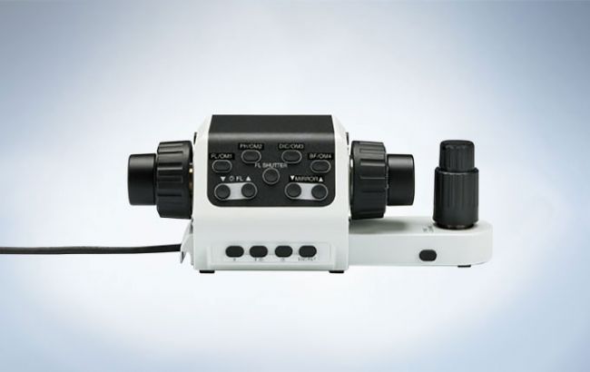
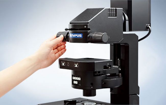
Intuitive and Ergonomic Microscope Control
Smart Control
Switch Observation Methods with a Single Touch
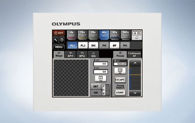
Olympus includes a touch panel controller with the IX83 that allows the user to easily configure all automated functions on the microscope. This includes advanced functions such as magnification-specific lamp intensity adjustment. The touch panel controller, used in combination with cellSens software, enables advanced customization.
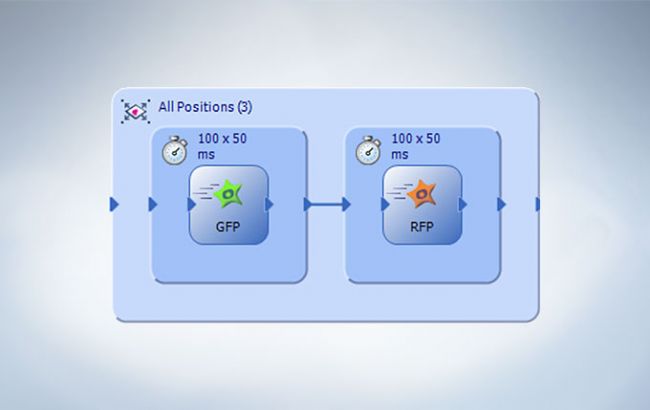
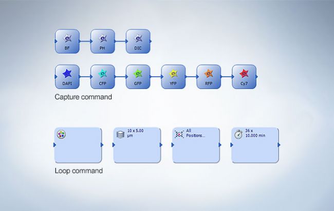
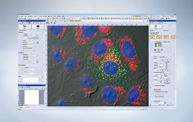
cellSens Graphical Experiment Manager (GEM)
GEM is a flexible drag-and-drop interface to build simple or complex experiments within cellSens software. Combine actions within specialized frames to dictate the order and priority of automation. Easily acquire multichannel, Z-stacks, and time-lapse imaging across one or more sample positions. Perform two-channel simultaneous imaging within GEM using the cellSens High-End Device solution. GEM permits users to interact with the system during long timelapse imaging without terminating the experiment.
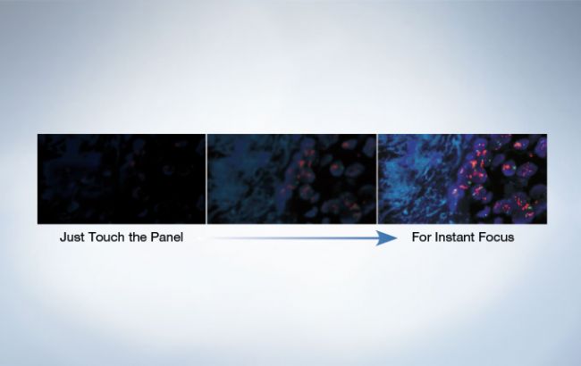
ZDC One-Shot Function Quickly Detects Focus, Even during High-Magnification Observation
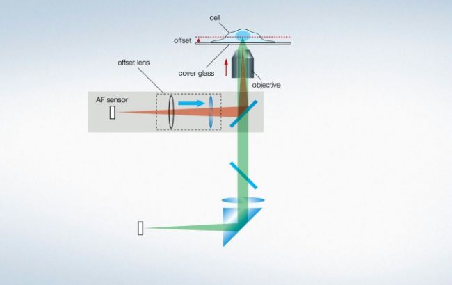
IX3-ZDC2 focus detection and tracking can be performed through the innovative touch panel controller, independent of software. There is also a focus search function supported by a cell-safe, near-infrared laser, allowing users to instantly focus on samples, even at high magnification.
Intuitive Microscope and XY Stage Controllers
The U-MCZ focus module and XY stage controller provide the familiarity of conventional handle operation for intuitive microscope control, even in a darkroom environment.
Stored Microscope Configurations (Olympus cellSens)
The system saves microscope configurations alongside image data by incorporating a readout of motorized and coded unit position. With this advanced system, a wide range of settings can be recalled to recreate the desired imaging conditions, thereby creating a highly reproducible and easy-to-use, high-end imaging system.
Operator-friendly Design
Reliable Tracking at High Magnification
The IX3-SVR manual stage features a reliable positioning system which enables the easy tracking of cells, even at high magnifications. The travel limits can be manipulated by the user to immobilize the stage and prevent inadvertent movement during critical operations. It is also possible to remove the 35mm dishes from the stage, place them in an incubator and return them to the exact location previously identified.
Easily Achieve Koehler Illumination
The condenser can be moved and easily reset to Koehler Illumination using the conveniently-located condenser lock and control knobs.
Catch Tray Minimizes Optical System Contamination
A catch tray under the nosepiece minimizes the possibility of contaminating and damaging the microscope with spilled liquids. This feature also simplifies maintenance.
Ideally Suited for Live Cell Imaging
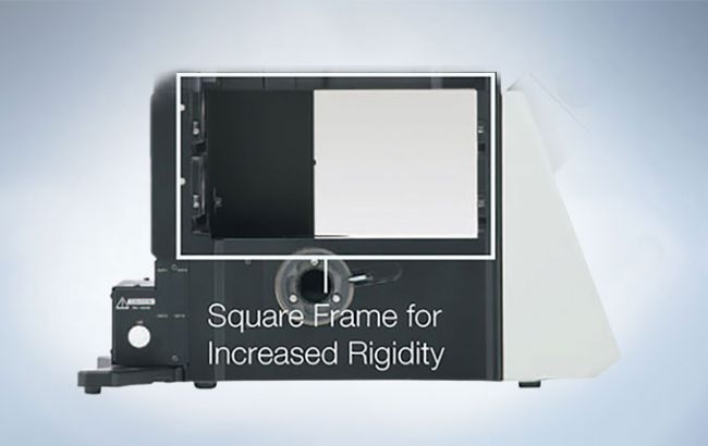
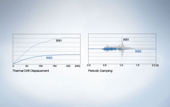
Easily capture dynamic cellular processes with the IX83, an imaging system designed for remarkable time-lapse imaging. With new frame architecture and focus drive design, the IX3 system offers enhanced rigidity that reduces the impact of vibration and temperature. It maintains desired positions along the X, Y, and Z axes to facilitate reliable time-lapse and multipoint imaging.When combined with the Olympus IX3-SSU ultrasonic stage and Z drift compensator (IX3-ZDC2) , the IX83 is perfectly suited for capturing high-precision, multipoint time-lapse images that are never out of focus or misaligned. Box-type and stage-top incubators are also available to maintain the viability of live cells during time-lapse experimentation.
Imaging Accuracy
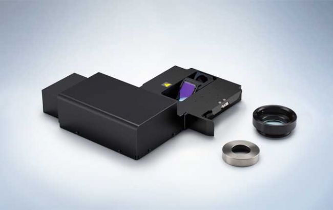
Z Drift Compensation System (IX3-ZDC2)
The new, fully motorized Olympus Z drift compensation module combats focal drift to keep the sample in sharp focus during long-term, time-lapse experiments. The improved IX3-ZDC2 is faster, more flexible and supports a larger range of objectives and sample vessels.
One-shot autofocus (AF) mode allows several focus positions to be set as desired for thicker samples, enabling efficient Z-stack acquisition in multi-position experiments. The enhanced continuous AF mode, now also supporting dry objectives, keeps the desired plane of observation precisely in focus, even during the addition of reagents or changes in room temperature.
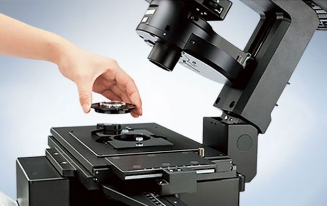
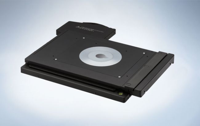
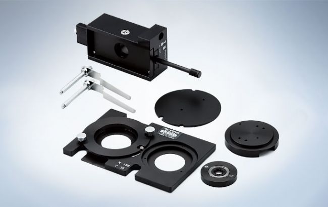
High-precision Ultrasonic Stage for IX3 (IX3-SSU)
With low thermal drift and high accuracy, the ultrasonic stage delivers excellent reliability for multi-image acquisition. Sample holders firmly secure slides or dishes to ensure accurate repeatability during high-magnification, multi-point observations.
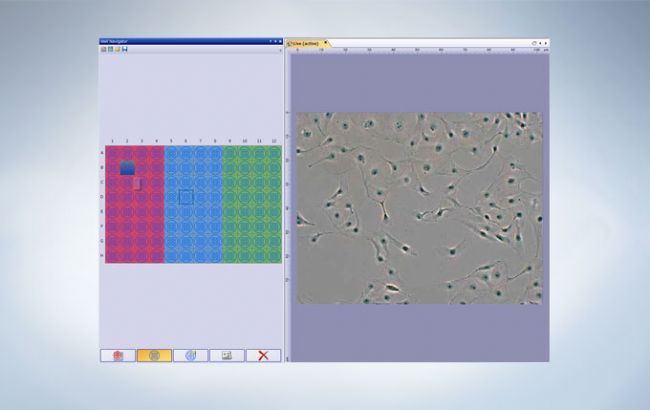
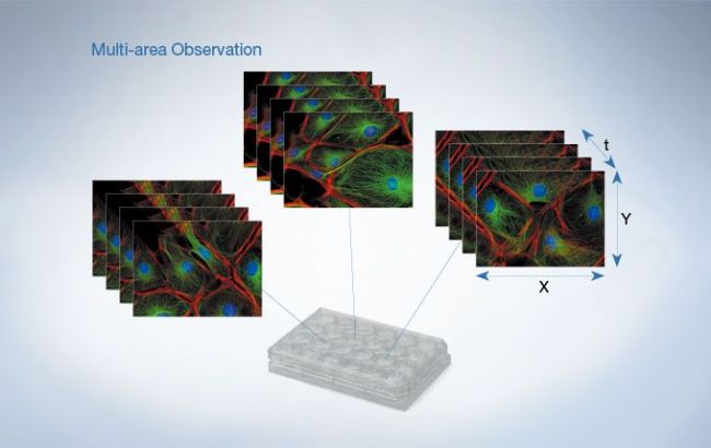
Efficient Multi-Image Acquisition with cellSens Software
The IX3 ultrasonic stage can be used with cellSens software to conduct multi-area time-lapse imaging with greater efficiency. Image cell dynamics at multiple points within each well and join adjacent fields to create wide area, time-lapse images and obtain a greater amount of data from the same experiment. The excellent accuracy and repeatability of the ultrasonic stage makes it easy to obtain seamless tiled images with cellSens software.
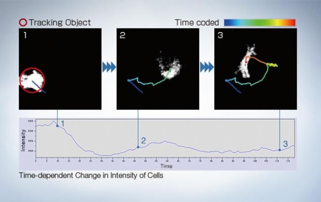
cellSens Object tracking
Detect, track, and analyze moving objects. cellSens Tracking provides an intuitive tool to quantify dynamic processes such as cell movement and division.
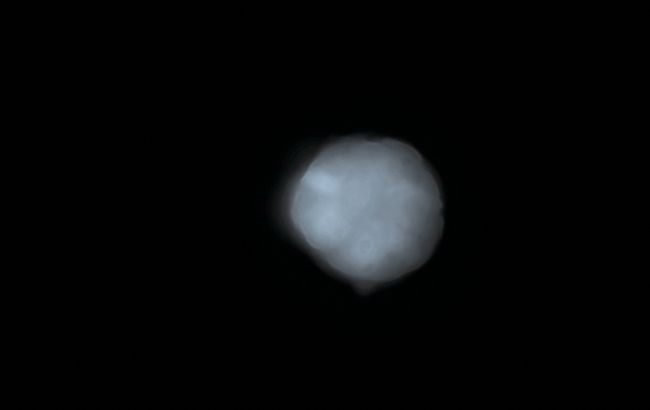
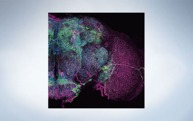
Bioluminescence of RA-induced differentiating cells at day 12 from Bmal1:luc stably transfected ES cells Image data courtesy of: Kazuhiro Yagita, M.D. Ph.D. Department of Physiology and Systems Bioscience, Kyoto Prefectural University of Medicine Reference: Proc Natl Acad Sci U S A. 107(8): 3846–3851(2010)
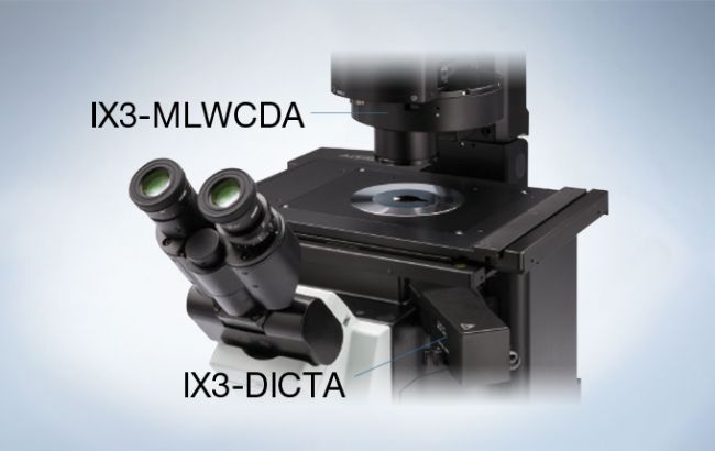
Motorized DIC Slider for IX83

The motorized IX3-DICTA allows users to engage and adjust DIC without disturbing the sample.
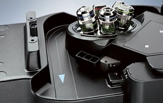
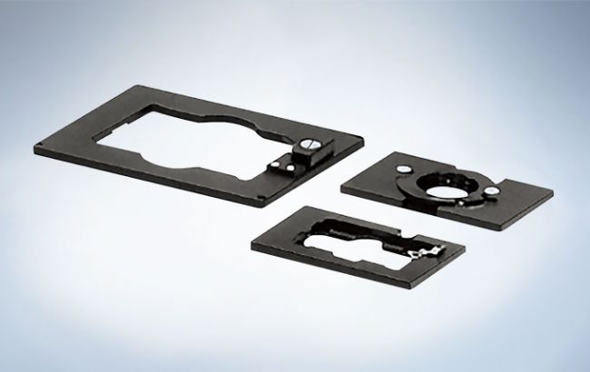
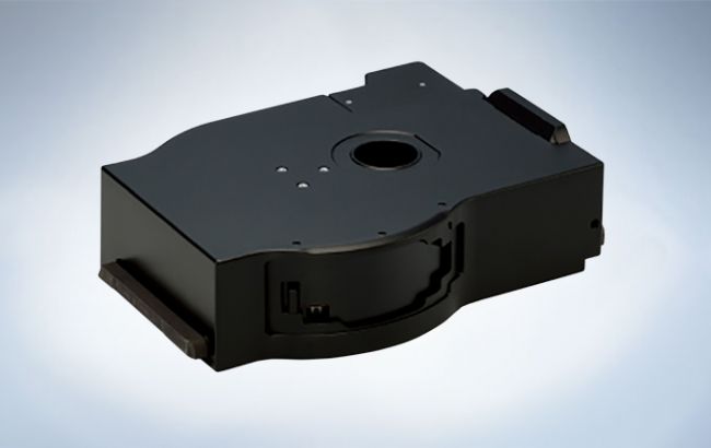
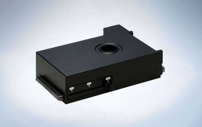
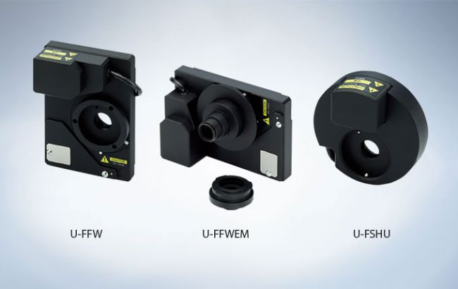
Interchangeable Modules Provide Flexible Imaging Options
A wide range of units are available for the Olympus IX3 microscope system, bringing enhanced usability to everything from casual observation to high-end imaging. Simple cassette-like insertion into the deck makes it easy to mount fluorescence mirror turrets, right side ports, mag changers, epi-illuminators and other units.
The large open frame allows a motorized emission filter wheel to be fitted within the infinity space of the microscope. This eliminates image shift between channels and allows the eyepieces to see what the camera detects.
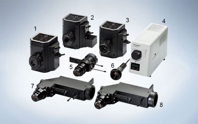
Deck Units/High Speed Units
Motorized Fluorescence Mirror Turret (IX3-RFACA)
A non-click turret that can be fitted with up to 8 mirror units and that delivers smooth, rapid cube rotation. The turret can accommodate both 25mm and 32mm diameter filter units, which can easily be inserted and removed without the need of tools.
Motorized/Manual Right Side Port with C-mount (IX3-RSPCA/IX3-RSPC)

The C-mount-equipped right side ports can be fitted with up to two mirror units, enabling advanced applications such as split imaging.
Coded Intermediate Magnification Changer (IX3-CAS)
Magnification can be easily changed between 1X, 1.6X and 2X by adjusting the lever. Since the system is coded, information on intermediate magnifications is saved with image data.
Motorized Fast Filter Wheels and Shutters
With Olympus filter wheels, the user can easily switch between filters in milliseconds. Olympus shutters operate even faster. The IX83 is capable of controlling up to six filter wheels and four shutters, thus enabling complex, multi-modal imaging.
Fluorescence System
Application-Specific Fluorescence Illuminators
There is an illuminator best-suited for each application. The L-shaped fluorescence illuminator with the Fly-Eye lens system (IX3-RFALFE) provides bright, even illumination without adjustment. The standard L-shaped fluorescence illuminator (IX3-RFAL) is cost-effective and is equipped with both field and aperture iris diaphragms. The straight fluorescence illuminator (IX3-RFA) is ideal for applications demanding intense excitation light. A wide range of light sources is also available, including counter top and frame-mounted units (mercury and xenon).
Long Life 130W Mercury Lamp (U-HGLGPS)
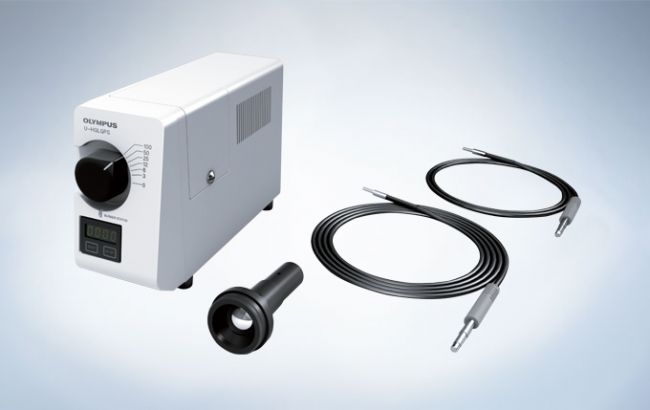
The U-HGLGPS fluorescence light source delivers bright, even illumination and features a long life, 2,000-hour mercury burner (average lifetime). The unit is maintenance-free and does not require centering adjustment. A liquid light guide prevents heat transfer to samples, thus allowing extended observations to be carried out without concern.
Measuring Excitation Light Intensity for High Reproducibility in Imaging Adapter for Excitation Irradiance Meter/IX3-EXMAD*
Olympus now offers an adapter for a power meter that can directly measure the excitation light intensity per unit surface area of the sample, as well as offering irradiance display software. Displays the measurement results on a monitor and records the data, eliminating The need for laborious calculations. This makes it possible to check the excitation light intensity before starting an experiment, enhancing the reliability of experiments. Data can also be easily shared.
*This equipment was based on the technical development at RIKEN BSI-Olympus Collaboration Center.
Total Internal Reflection Fluorescence (TIRF) Microscopy
Total internal reflection fluorescence (TIRF) microscopy has become firmly established over the last few years as a key technique in the investigation of molecular interactions at or near the cell surface. Olympus is experienced in providing advanced TIRF solutions.
cellTIRF-4Line system
The cellTIRF-4Line system is easy to use for TIRF, single molecule localization microscopy and FRAP for a range of experimental protocols.
cellTIRF-1Line system
A cost-efficient device with motorized TIRF angle control, the single-line cellTIRF-1Line system illuminator can also be equipped with laser combiners.
-
Olympus is one of the world’s leading manufacturers of professional opto-digital products for medicine, science and industry. As a result, Olympus provides a comprehensive range of solutions. From microscopes for training and routine tasks to high-end system solutions in the fields of life science, there is a system for every need. The product line is complemented by innovative laboratory equipment for cellular research applications and the new all-in-one microscopes that offer user engagement at all levels.
| Request Information |
| Related News |
| Other Products |