

- Research Macro Zoom Microscope

- Product Detail
- Company Profile
Fluorescence techniques are ideal for the observation of whole organisms because they are non-destructive and can be performed over long periods. The perfect microscope for fluorescence observation of intact organisms must combine maximum detection efficiency from macro-to-micro magnifications with a high zoom and high NA for resolution of fine details. The MVX10 MacroView brings these factors together with low autofluorescence and other unique features to bridge the gap between macro and micro observation and providing unprecedented brightness, resolution and precision to the imaging system. The MVX10 provides a solution to researchers interested in the impact of gene expression and protein function at the cellular level as well as within whole tissues, organs and organisms.
Bright and High-Precision Macro Fluorescence
Single-Zoom Concept for Bright Fluorescence

The MVX10 MacroView, employs a single-zoom optical path with a large diameter, which is optimized to collect light with revolutionary efficiency and resolution at all magnifications. From fluorescent observation of whole organisms, such as zebrafish, at low magnification to the detailed observation of gene expression at the cellular level at high magnification, the MVX10 features a unique pupil division mechanism in the light-path to mimic the effect of a stereo microscope.
Dedicated to Advanced Fluorescence Imaging
All optical components contribute to the phenomenal fluorescence performance of the MVX10. Incorporating the latest technologies and materials, the objectives and HQ filters produce minimal autofluorescence. Together with high numerical apertures, this results in a maximal S/N ratio for excellent contrast of faint fluorescence signals. All filter cubes absorb stray light and the aspherical fluorescence collector bundles the light for minimum intensity loss.
Seamless Observation from 4x to 125x

With the parfocal objectives and two-position revolving nosepiece, switching between objectives is very quick and easy, requiring little refocusing. Therefore, with the 0.63x and 2x objectives, the uninterrupted magnification range is 4x to 125x (eye observation with 10x eyepieces). This in combination with the 2x magnification change slider, increases effective magnification to 250X.
High Optical Performance on Every Application
Unique Objective Line
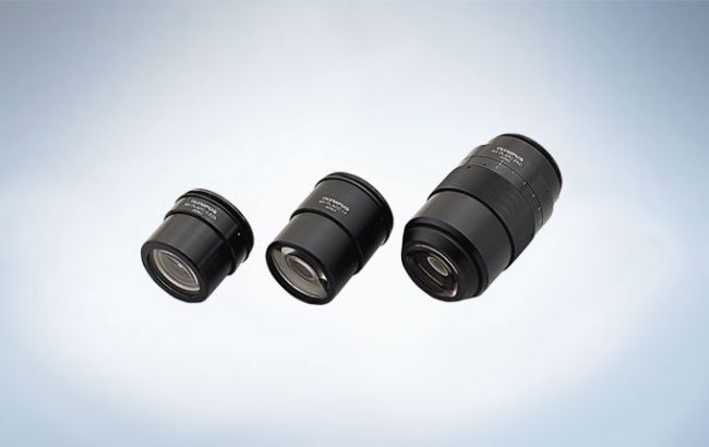
While MVX10 provides the same working distance and large field of view as a stereo microscope, it also benefits from having increased numerical (NA), leading to greater resolution. Specially designed 0.63x, 1x, and 2x planapochromatic objectives for the MVX10 produce high quality images. All three objectives are pupil-corrected for outstanding image flatness and show high transmission to NIR and excellent chromatic aberration correction. This provides great flexibility for efficient, fast and precise fluorescence observation, screening and imaging — from low to high magnification over time.
Ideal for Fast Moving Specimens
The 0.63x objective has a maximum field of view of 55mm, making it easy to track fast-moving specimens over time. With its exceptionally high NA of 0.15, fluorescence from large objects, such as whole embryos, can be viewed with outstanding brightness at all magnifications.
Optimized for Sample in Medium
The 2.0x objective available for the MVX10 has the highest NA at 0.5. This plan apochromat is perfect for working with samples in media. Its correction ring enables fine adjustment of the lenses to correct for mismatches caused by the different refractive indices of the vessel and medium. This helps to reach the maximum resolution of C.elegans vessels and Zebrafish in media.
Contribution to Work Efficiency
Ample Working Space with High Resolution

This makes fluorescence screening and verification of gene expression especially efficient, improves speed and precision, reduces judgment errors, and eliminates the need to switch back and forth between a stereomicroscope and inverted microscope.
Minimum Sample Damage
The peerless NA and S/N ratios of the optical components mean that specimens can be exposed to the excitation light for shorter periods. This is also true at near-infrared wavelengths, where the MVX10 has excellent transmission properties throughout the entire spectrum, minimizing damage to the specimen.
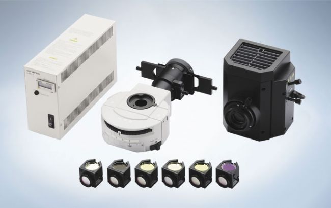
Accessories
Unique Color & Monochrome Camera

The DP80 uniquely combines both color and monochrome sensors within the same housing, providing high resolution with excellent photon detection. With versatile functionality, addition of the cellSens control software enables rapid, automatic exchange between monochrome and color, without mechanically switching the microscope optical path. Additionally, the DP80 has the capacity to overlay images from the two sensors with accurate pixel-to-pixel correspondence, presenting significant opportunities for efficient and accurate joint color and fluorescent imaging in both research and clinical environments.
Click here for more information about Olympus cellSens software
Click here for more information about Olympus DP80 digital camera
Choice of Optimum Illumination Bases
SZX2-ILLB
The high-level transmitted-light stand for brightfield and oblique illumination.
Its high and low contrast selection provides clear, effective contrast and illumination.
Light intensity and color temperature are easily adjusted on 6 V, 30 W halogen lamp.
Used with a high-magnification objective, it permits the observation of extremely small details on highly contrasted structures; which particularly effective for C. elegans, oocytes, and embryos.
SZX2-ILLD
Transmitted-light stand for brightfield and darkfield. It has a built-in 6 V, 30 W halogen lamp and can be switched between brightfield and darkfield illumination, reducing background noise and improving contrast to resolve even the finest details.
SZX2-STL
The SZX2-STL stand a stable, broad working space for observing large specimens. Attaching the Motorized Focus Unit (SZX2-FOA) creates a more comfortable work environment.


-
Olympus is one of the world’s leading manufacturers of professional opto-digital products for medicine, science and industry. As a result, Olympus provides a comprehensive range of solutions. From microscopes for training and routine tasks to high-end system solutions in the fields of life science, there is a system for every need. The product line is complemented by innovative laboratory equipment for cellular research applications and the new all-in-one microscopes that offer user engagement at all levels.
| Request Information |
| Other Products |
| Recently viewed products |