The deduced 673-amino acid protein contains a putative hydrophobic signal sequence, 10 tandem arrays of a leucine-rich repeat, an epidermal growth factor-like domain, a fibronectin type III-like domain, and a short intracellular region. In situ hybridization detected mouse vasorin in the tunica media of the proximal ascending aorta, descending thoracic aorta, abdominal aorta, and coronary arteries. In mouse kidney, vasorin was expressed in interstitial cells. expression of vasorin in aortas increased gradually in parallel with the differentiation of vascular smooth muscle cells beginning at embryonic day 10.5. Vasorin expressed by transfected Chinese hamster ovary cells showed an apparent molecular mass of 110 kD, which shifted following N-glycosidase treatment. Vasorin was expressed on the cell surface and was secreted into the culture medium.
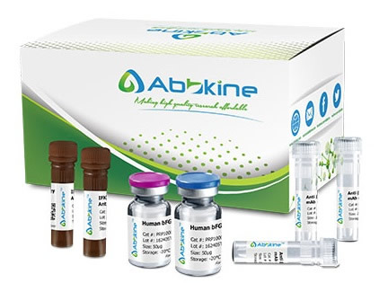
Human Vasorin (VASN) ELISA Kit employs a two-site sandwich ELISA to quantitate VASN in samples. An antibody specific for VASN has been pre-coated onto a microplate. Standards and samples are pipetted into the wells and anyVASN present is bound by the immobilized antibody. After removing any unbound substances, a biotin-conjugated antibody specific for VASN is added to the wells. After washing, Streptavidin conjugated Horseradish Peroxidase (HRP) is added to the wells. Following a wash to remove any unbound avidin-enzyme reagent, a substrate solution is added to the wells and color develops in proportion to the amount of VASN bound in the initial step. The color development is stopped and the intensity of the color is measured.
Human Vasorin (VASN) ELISA Kit listed herein is for research use only and is not intended for use in human or clinical diagnosis. Suggested applications of our products are not recommendations to use our products in violation of any patent or as a license. We cannot be responsible for patent infringements or other violations that may occur with the use of this product.
bio-equip.cn


