Phosvitin, a highly phosphorylated glycoprotein, represents the major fraction of hen egg yolk phosphoproteins. Circular dichroism, Fourier transform infrared spectroscopy, and Fourier transform infrared photoacoustic and fluorescence spectroscopic methods were employed to determine the secondary structure of the protein in both the solid and solution phases. This was supplemented by a Chou-Fasman type of predictive algorithm for the first 25 residues at the N terminus of the dephosphorylated protein. A three-compartment model consisting of alpha-helical, beta-sheet, and beta-turn components with beta-turns occurring at the interface between alpha-helical and beta-sheet regions in the proximity of O-phosphoserine residues is suggested from the combined analyses. Beta-sheets appear to be the dominant secondary structural component in phosvitin in the solid and solution phases.
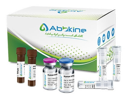
Fish Phosvitin (PV) ELISA Kit employs a two-site sandwich ELISA to quantitate PV in samples. An antibody specific for PV has been pre-coated onto a microplate. Standards and samples are pipetted into the wells and anyPV present is bound by the immobilized antibody. After removing any unbound substances, a biotin-conjugated antibody specific for PV is added to the wells. After washing, Streptavidin conjugated Horseradish Peroxidase (HRP) is added to the wells. Following a wash to remove any unbound avidin-enzyme reagent, a substrate solution is added to the wells and color develops in proportion to the amount of PV bound in the initial step. The color development is stopped and the intensity of the color is measured.
Fish Phosvitin (PV) ELISA Kit listed herein is for research use only and is not intended for use in human or clinical diagnosis. Suggested applications of our products are not recommendations to use our products in violation of any patent or as a license. We cannot be responsible for patent infringements or other violations that may occur with the use of this product.
bio-equip.cn


