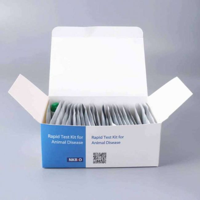
African Swine Fever Virus Fluorescent PCR Detection Kit
(Blood / Blood Cell / Lymph Nodes / Spleen / Tonsils)

INTENDED USE
For the detection of African Swine Fever Virus nucleic acid in blood, blood cell, lymph nodes, spleen and tonsils.
Naked eyes interpretation for Qualitative Results , Quantitative Analysis with machine.
KIT CONTENTS
|
Composition
|
Components
|
Specification
|
|
Box A
|
Buffer A
|
25 mL/Bottle×1 Bottle
|
|
Buffer B
|
60 mL/Bottle×1 Bottle
|
|
Buffer C
|
1250μL/Tube×2 Tubes
|
|
DNase-Free Adsorption Column and Collection Tube
|
50 Kits
|
|
Technical Manual:
|
1 Copy/Kit
|
|
Box B
|
Fluorescent PCR Reaction Solution
|
1000μL/Tube×1 Tube
|
|
Negative Control
|
1000μL/ Tube×1 Tube
|
|
Positive Control
|
1000μL/ Tube×1 Tube
|
STORAGE AND STABILITY
1. Box A store at room temperature (2-30℃).
2. Box B store at -20℃.
3. The shelf life is 12 months.
TEST PROCUDURE
Viral DNA extraction (using Box A)
(1)The sum of the number of samples to be tested is denoted by “n”, take n sterilized 1.5mL centrifuge tubes and number each tube.
(2)Add Buffer A 500μL per tube.
(3)Add 200μL of processed samples to each tube, mix thoroughly and leave at room temperature for 10 min.
(4)Take the same amount of DNase-Free adsorption columns and collection tubes as the above centrifuge tubes and number them. Transfer the solution in the centrifuge tube to the DNase-Free adsorption column (to prevent clogging of the adsorption column, do not absorb suspended impurities).
(5)Centrifugation at 13000 r/min for 30s at room temperature
(6)Discard the liquid in the collection tube and place the adsorption column back into the collection tube.
(7)Add 600μL Buffer B to the adsorption columm, centrifuge at 13000 r/min for 30s.
(8)Discard the liquid in the collection tube and place the adsorption column back into the collection tube.
(9)Repeat procedure (7), (8).
(10)Centrifuge the column at 13000 r/min for 2 min to remove the residual liquid.
(11)Transfer each adsorption column into a new 1.5mL centrifuge tube, and add 50μL of Buffer C to the center of the column. stand at room temperature for 1 min, centrifuge at 13000 r/min for 30 s. The liquid in the centrifuge tube is the template DNA. The obtained DNA solution should be stored on the ice for later use (Note: The extracted DNA should be PCR amplified within 2h. For long-term storage, place in -80℃ refrigerator to avoid repeated freezing and thawing).
Fluorescent PCR Amplification (using Box B)
(1)Remove the fluorescent PCR reaction solution from the kit, melt it at room temperature, and centrifuge at 2000 r/min for 5s. Set the required number of PCR tubes to “n” (n= sample number+1 tube negative control +1 tube positive control), and dispense 20μL of PCR reaction solution into each PCR tube.
(2)Respectively Add 5μL each of the positive control, negative control and DNA solution prepared in 1.3.1(11) to the above PCR tubes, tighten the cap and centrifuge at 500 r/min for 30s.
(3)Place the PCR tubes into the fluorescent PCR detector after sample addition and mark it. Reaction parameter setting:
Stage1, pre-denaturation 95℃/3 min;
Stage2, 95℃/15s,60℃/35s,45 cycles total. Fluorescence collection is performed at 60°C extension for each cycle of the Stage2.
READING RESULTS
| |
FAM |
HEX |
Statement |
| ASFV-Positive |
≤38 and S-shaped amplification curve |
≤38 and S-shaped amplification curve |
Strong positive sample HEX channel occasionally not amplified |
| ASFV-Negative |
>38or no CT value |
≤38 and S-shaped amplification curve |
HEX channel amplified, FAM not |
| Invalid |
None |
None |
No amplification curves for HEX and FAM |


