

- Virtual Slide Microscope

- Product Detail
- Company Profile
The VS120-S6-W slide loader system allows for manually loading one and six standard slides, respectively, along with any associated meta-data. Designed for high throughput research and pathology, the VS120-L100-W system features a highly dependable, robustly designed slide loader for up to 100 slides.
The VS120 is not for clinical diagnostic use.
Ideal for Research Facilities and Digital Learning
State-of-the-art Research Tool for Brightfield and Fluorescence
The VS120 not only creates high resolution brightfield images, but also can scan in full multi-fluorescence mode. Utilizing virtual microscopy for fluorescence imaging helps to minimize problems associated with damaging and fading of sensitive fluorescence samples.
An innovative new algorithm makes image stitching more precise than ever, enabling high-level accuracy that can be applied from small animal brain slices to large specimens. The VS120 also switches seamlessly between micro and macro observation to enable swift viewing of regions of interest and overall structures alike.
Advanced Medical Education and Collaboration
The VS120 allows multiple viewers to study virtual slide specimens simultaneously via simple server access, regardless of time and location - providing an ideal solution for medical instruction, Q&A session and remote collaboration.
Remote Conferencing and Consultation

Virtual slides can be archived to a database, enabling network-based remote retrieval at any time through the Olympus Net Image Server SQL. Images are stored at high resolution, and multiple clients can review and even synchronize elements such as specific observation areas to facilitate efficient review and discussion.
High-detail, High-precision, Rapid Scanning
Always in Focus with the VS120 Focus Map
To optimize scan speed and efficiency, the VS-ASW generates a focus map using the software-based autofocus function. This allows the optimal Z-position to be determined and saved on various parts of the sample, permitting a height profile of the sample to be compiled before detailed scan acquisition. Different maps are used depending on the type of sample being scanned, providing the best solution whatever the specimen. Numerous coordinates are selected to create the map, which gives the system an instant focus reference when scanning the slide.
Wide Range of Objectives from 2x to 100x

The VS120 comes standard with Olympus UPLSAPO 2x, 10x, 20x and 40x objectives, allowing users to choose the objective most suitable for their research needs. Automatic specimen recognition capability limits scanning to the specimen area, with high-level color fidelity and image quality. Additionally, UPLSAPO 60x and 100x oil immersion objectives are provided as an option for a VS120-S6-W configuration.
Virtual-Z with 3D Virtual Slide Production
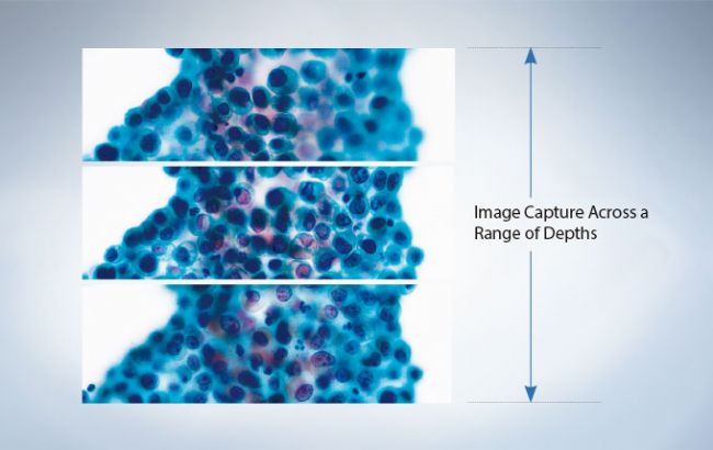
The VS120 can scan multiple large specimens to 31 Z-planes. Multi-plane virtual slides can then be produced by simply selecting attributes such as depth for multiple areas, range, number of planes, and magnification within the virtual-Z scan mode.Virtual-Z scanning also allows the user to adjust the image to the specific desired depth with a simple scroll of the mouse - a function particularly advantageous for the viewing of thicker specimens, such as cell clusters or cranial nerves.
Accurate Image Stitching, Regardless of Specimen Quality

The VS120 minimizes stitching errors by automatically recognizing only where the specimen exists and then integrating images of consecutive areas.A newly developed algorithm makes image stitching with the VS120 so precise, it can even capture quality composite images across large and uneven specimen regions.
High-resolution, High-sensitivity Virtual Fluorescent Slides
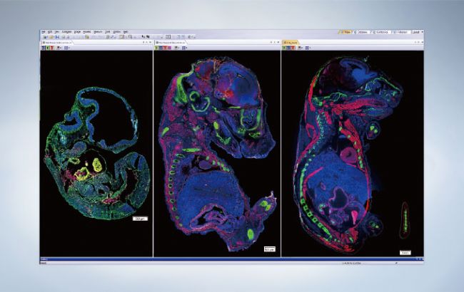
High-speed filter wheels of the fluorescent unit can be installed on both the excitation and observation side, enabling the swift production of fluorescent virtual slides with high-level definition and resolution. Multi-colored virtual slides also can be prepared for long-term observation, negating concerns over fading, discoloration, and degradation.
Automation Enhances Laboratory Efficiency
An optional automated slide loader with a capacity to hold 100 slides adds efficiency to laboratories with high-throughput requirements. Furthermore, specimen information can be automatically read using 1D and 2D barcode scanners, making it easier to store and organize information.
Intuitive slide scanning workflow and data review
Full Slide Scanning in Three Clicks of the Mouse
The VS120 system components are designed to interact seamlessly, producing a fully automated high-speed scanning and reviewing system with excellent flexibility and simple operation.
Efficient and robust VS-ASW acquisition software is provided with all VS120 Virtual Slide systems. The software facilitates the slide scanning process through an intuitive slide scan wizard, allowing the user to set up simple brightfield scans in just 3 mouse clicks. Several pre-defined modes facilitate ease-of-use for entry-level users, while the Expert setting puts experienced users in complete control of the entire scanning process. Therefore, different scan settings can be assigned to individual slides, saving significant scanning time, as well as offering the ability to only scan areas of interest, reducing data file sizes.
Instantly Get to the Desired Scanning Area
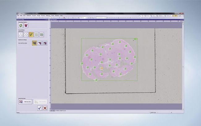
As the VS120 displays the scan area and focus mapping settings on the same screen, the desired scan area can be selected in an instant - bringing greater efficiency to the overall workflow avoiding the need to switch screens.
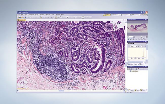
View Full and Magnified Images on the Same Screen
Both the whole slide and zoomed-in region can be displayed on the same screen, making it easy to pinpoint the specific location on the larger image.
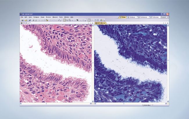
Innovative Synchronizing Enables Comparative Viewing of Different Stains
Analysis of multiple virtual slides prepared from the same specimen is made easy through the ability to align them on the monitor with positions and magnifications interlinked.
Save Voice Annotation Data
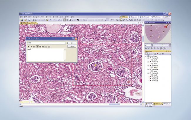
An innovative annotation function allows the user to save and link text and voice data to specific regions of interest on the slide.
Capture Clear Images with All-in-Focus Imaging for Fluorescence
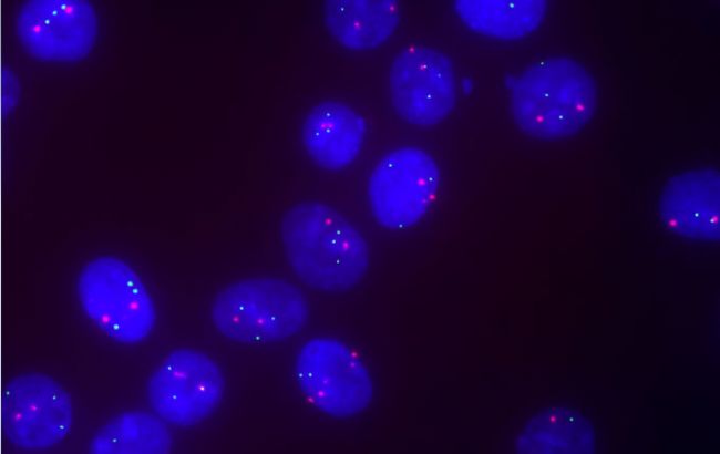
With the aid of virtual-Z scan mode, in-focus image data at different focal planes throughout the whole Z-direction can be captured and on-the-fly be merged using the Extended Focal Imaging or Z-projection function to generate an all-in-focus image.
This allows to visualize and observe fluorescence in irregularly shaped or thick samples, such as FISH samples.
Powerful data management with NetImage Server SQL (optional)
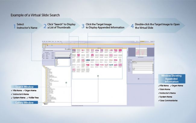
Fast Data Access
Virtual slides are easily found by using keywords through the folder tree. Simply double-clicking on the corresponding thumbnail image opens the desired virtual slide in a new window.
SQL-based Net Image Server
Building on the importance of high throughput, the optional, versatile Net Image Server SQL client server database, allows users to manage any image in a simple and convenient way. The database software allows to store images and share image data via web, such that the virtual slide images from VS120 can easily be released and shared to a wide audience. Access to the image data can be controlled via individual access rights.
Attach Metadata to Virtual Slides
The VS120 provides editable metadata fields that can be used to store data such as tissue name, staining method, organ name, system, instructor’s name and other keywords. Such information appended to slides, can assist greatly in an educational setting.
Batch Management of Digital Content

Offering functionality beyond virtual slides, the VS120 allows a wide range of image data to be archived to a database in both JPEG and TIFF formats, including macro images captured by other devices such as endoscopic images, X-ray images and electrocardiograms. Users are also able to save Microsoft Word, Excel and PowerPoint documents to the database.
Remote Access with Virtual Slide Viewers
OlyVIA Desktop
OlyVIA Desktop is a dedicated viewer software for Windows PCs. It is especially suitable for the viewing of VS120 virtual slide images. Images can be opened from local or network storage. Images that have been saved on Net Image Server (NIS) SQL can be viewed over the internet. OlyVIA Desktop supports image annotations and allows participation in conferences hosted by users with NIS SQL. Software Free OlyVIA 2.9 Viewer Download the free viewer software for virtual slide images via Software Downloads page.
OlyVIA Mobile
The Olympus OlyVIA mobile App, is a viewer for microscopic images which are located on the Olympus Net Image Server, NIS SQL. It allows fast and easy remote viewing anywhere at any time even of high resolution digital slides captured with the Olympus VS120 slide scanning system. Download iPad app via AppStore;
Web Access
A platform independent access to virtual digital slides can be provided with an Internet browser. No additional software installation is required for researchers to access images that have been saved on the Olympus Net Image Server, NIS SQL via internet.
-
Olympus is one of the world’s leading manufacturers of professional opto-digital products for medicine, science and industry. As a result, Olympus provides a comprehensive range of solutions. From microscopes for training and routine tasks to high-end system solutions in the fields of life science, there is a system for every need. The product line is complemented by innovative laboratory equipment for cellular research applications and the new all-in-one microscopes that offer user engagement at all levels.
| Request Information |
| Other Products |
| Related Products |
| Recently viewed products |