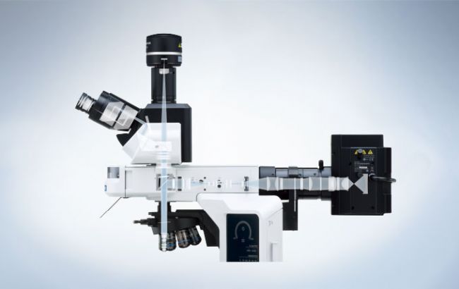
The BX53 microscope’s ergonomic design helps you stay comfortable during extended periods of use while the intuitive control layout enables fast, efficient observation and imaging. Optimized for laboratory applications, true color LED illumination has a high luminosity and color rendering index so you can see samples in true-to-life colors.
High Luminosity True Color LED Illumination
With an LED illuminator equivalent in brightness to a 100 W halogen lamp, the BX53 microscope delivers outstanding brightness that's ideal for teaching and polarized light applications.
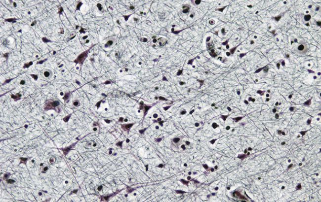
Designed for Pathology and Cytology
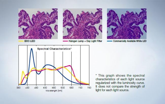
- Clearly view purple, cyan, and pink dyes
- Consistent color temperature; don’t waste time adjusting color filters
- 50,000 hour use life
Maintain Brightness when Changing Magnifications
The Light Intensity Manager eliminates the step of adjusting lamp brightness when changing magnification, so you can complete your observations quickly and with reduced eye strain.
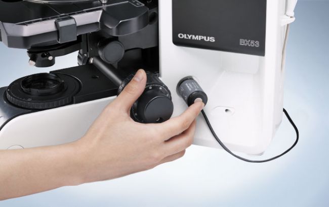
Quick Magnification Change with Motorized Functionality
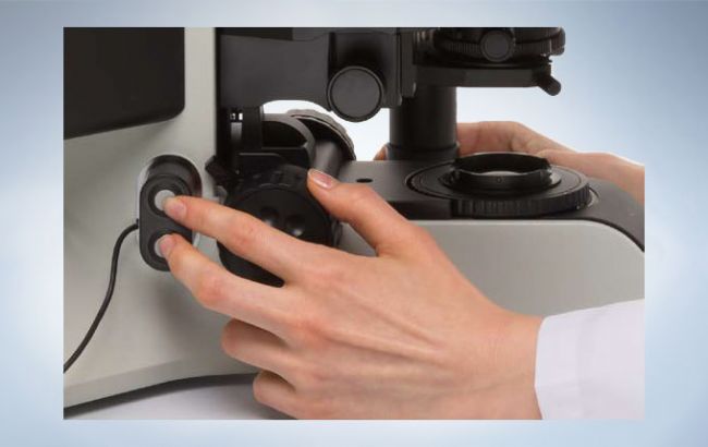
Easily change objectives with a motorized nosepiece. Position the hand switch near the focus handle to control the nosepiece without taking your eyes off the specimen.
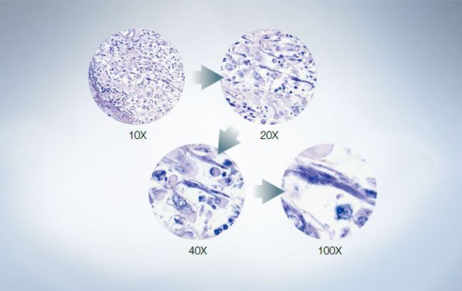
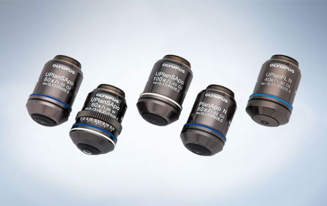
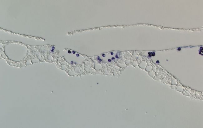
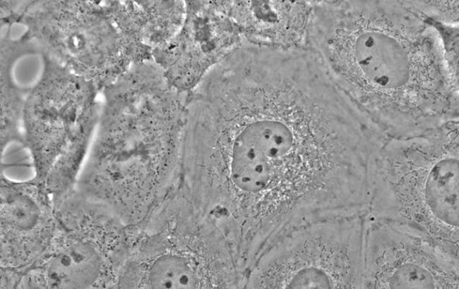
Suitable for Cellular Tissue Observation / LPLN40X
This objective is ideal for imaging thick, clear samples, even at 40X magnification.The LPLN40X is equipped with a correction collar so users can adjust the spherical aberration caused by differences in cover glass thickness to get clear images.
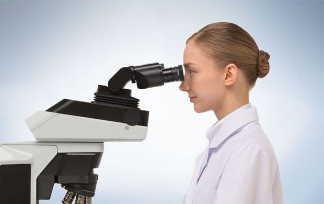
Stay Comfortable While You Work
The most ergonomic position differs from person to person, so it’s important that the microscope can accommodate each user down to the millimeter. Our ergonomic observation tubes enable you to adjust the inclination angle, extend the tube, and adjust the tube’s height.
Excellent Ergonomic Tube
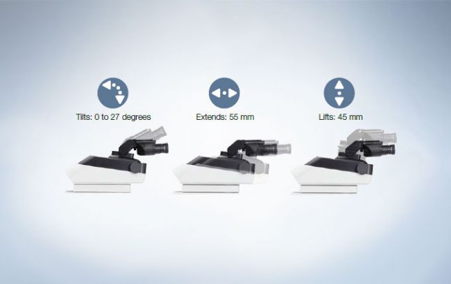
Our most ergonomic option, the excellent ergonomic tube, moves up and down, tilts, and extends forward and back so you can move it closer to you. It’s great for labs where multiple users share a microscope since each can adjust it to accommodate their height and posture.
Tilting Trinocular Tube

The tilting trinocular tube is designed for users who want the flexibility of
an ergonomic component but need to attach a camera to their
microscope. Attach the optical path switch to either side of the tube to accommodate both left- and right-handed users.
Tilting Binocular Tubes that Meet Your Needs
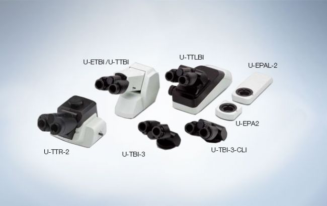
Choose from cost-effective models to tubes for erect image observation and eyepoint adjusters that accommodate users of different heights.
Comfortable, Easy-to-Use Stage
Rackless Stage
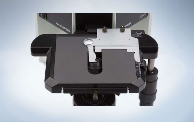
- Rackless, wire-driven design with no teeth in the gear helps minimize injuries
- Mechanical stages are coated with a durable ceramic for abrasion resistance
Keep Your Hands on the Desk
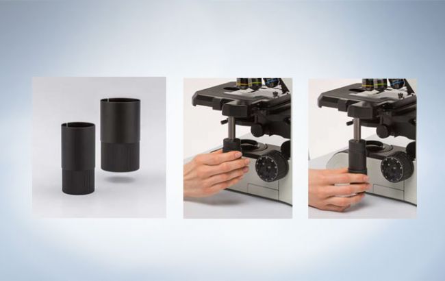
- Keep your arms resting on the desk with the stage handle extender
- Mount a rubber cap to the handle to control the stage with minimal force
cellSens Real-time Panoramic Imaging
Create stitched images in real-time with the Manual Process solution. Manual Process Conrtrol is available as an option for cellSens Standard software and included within cellSens Dimension software.
Efficient Image Capture
Easily Acquire High-Quality Images with cellSens Software
- Simple layout improves the efficiency of your workflow, from novice to expert
- All image acquisition functions are easily accessible; users with little training can obtain excellent results
Save Microscopy Data via Coded Units
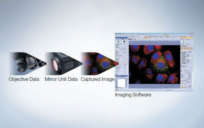
BX3 series microscopes include an optional coded nosepiece and mirror turret modules.
- Automatically record and share microscope magnification and setting information for post-imaging treatments
- The metadata is automatically sent to Olympus’ cellSens software packages, helping minimize mistakes and scaling errors
Hand Switch for Image Capture
Mount the remote exposure knob on either side of the microscope to acquire images at the touch of a button without having to look at the monitor or use the mouse.
Capture Digital Images without Using a PC via the DP22 Camera
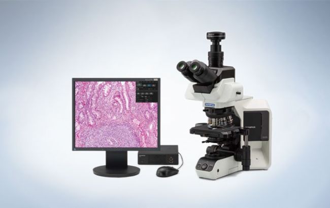
- Observe, measure, and acquire images without using a PC
- Precise color reproduction and smooth live images
- Display your specimen on a monitor and capture images for reports and conferences
Advanced Sensitivity in Fluorescence Imaging
The BX53 microscope delivers fluorescence images with a high signal-to-noise ratio (S/N) for bright colors and a dark background. High transmission objectives, mirror units, and a fly-eye lens system create uniform illumination and detection.
Fluorescence Illuminators with a Fly-Eye Lens

- Even illumination across the field of view
- Homogenous illumination across the entire wavelength spectrum
- Simpler burner alignment
Integrated Flexibility with an 8-Position Fluorescence Illuminator
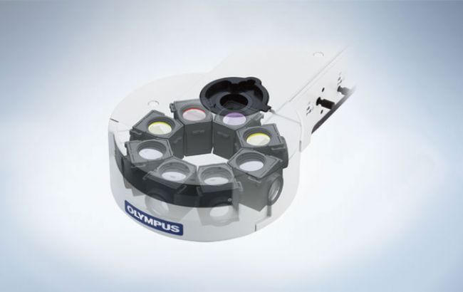
- No need to replace the mirror units for multicolor or FISH applications
- If you do need to exchange the mirror units, it’s easy to do and no tools are required
cellSens Capture Multidimensional Images
The Process Manager makes it easy to capture multicolored and multidimensional images with just a couple of clicks.
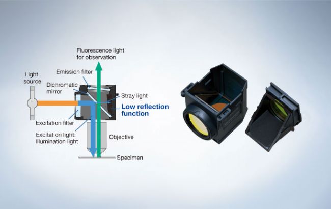
Fluorescence Mirror Units with Advanced Coatings and Stray Light Reduction
- UIS2 fluorescent mirror cube optimized for fluorescence imaging
- High-quality coatings provide excellent transmission and steep cut-off slopes
- Interior surfaces eliminate over 99% of stray light for high sensitivity and excellent color separation
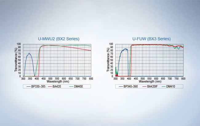
High Transmission with Reduced Autofluorescence
- UIS2 high NA objective lenses are corrected for chromatic aberrations to deliver high resolution, even from faint fluorescence signals
- Advanced UW multi-coating technology reduces autofluorescence and improves the signal-to-noise ratio
- UW multi-coatings also yield a flat, high transmission over a wide wavelength range for high performance in research that uses different fluorochromes
Condenser Designed to Reduce Back-Reflections
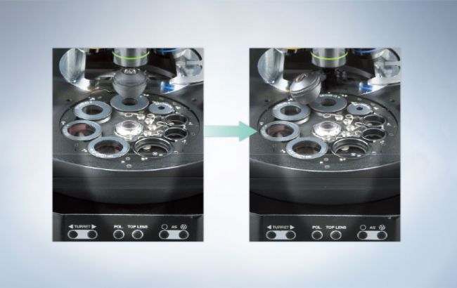
The motorized universal condenser is designed to reduce back-reflections and autofluorescence by swinging its top lens out, automatically closing its diaphragm to the minimum, and locating the wheel in between two positions during fluorescence imaging.
Low Autofluorescence Immersion Oil

- Reduces noise for an improved signal-to-noise ratio; helpful in quantitative observation of single molecule fluorescence
- Resistance to crystallization enables the oil to be used over a long period of time
- The oil’s refraction index is the same as other Olympus products for easy integration
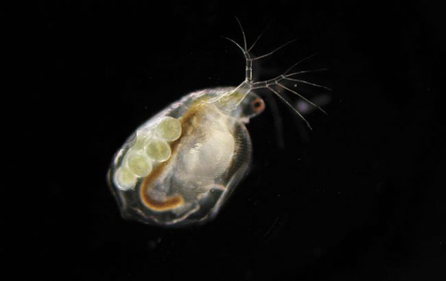
Renowned Optical Performance
Biological samples often don’t possess inherent contrast, such as color variations, when observed using brightfield illumination. As a result, different optical contrast methods and sample contrast methods have been developed. Whatever the source of contrast, our BX3 series microscopes and UIS2 optical components provide sharp, clear images in any contrast method.


Designed to Meet Expanding Needs
Research microscopy is about more than just the microscope — each investigation requires a unique setup. Our BX3 series microscopes are customizable with modular hardware and software. Choose the components you need for your research application, or start with a base system that can grow with your research.
Bright Images in Multi-Head Configurations
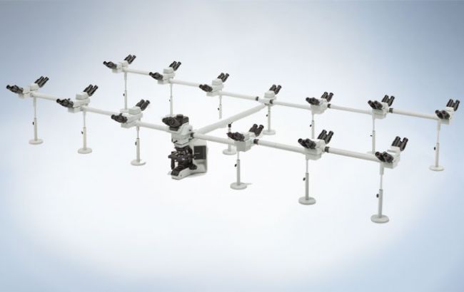
Multihead discussion systems are essential for training and education. With the BX53 microscope's high luminosity LED illumination, up to 26 participants can view clear, bright images.
Digital Imaging to Fulfill Diverse Needs
Tailor the system to your application, from advanced research work to stand-alone models for conferencing. Our full line of digital cameras and cellSens imaging software help ensure fluorescence imaging with a high S/N ratio.
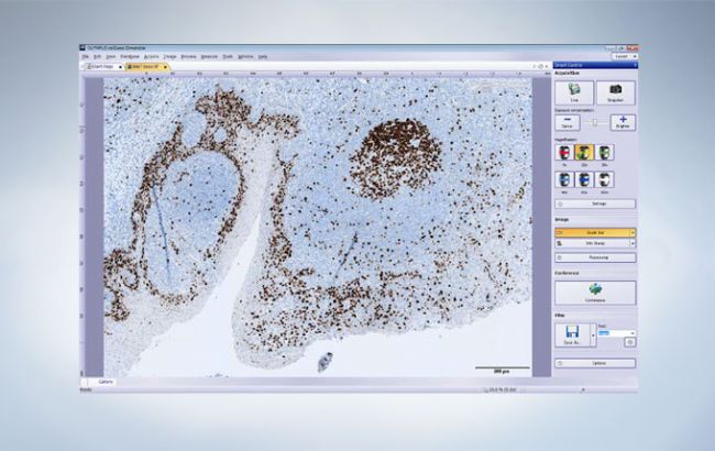
cellSens Simple Layout
Novice and expert users will simultaneously benefit from the "Simple Layout," an interface designed for a clinical research workflow. Acquire, annotate, share, and save your images using the intuitive Smooth Control Tool Window. Built-in measurement tools display only when required, reducing software clutter and minimizing distraction.
cellSens Conference Mode
Use Conference mode to fill the screen with live or static images for presentation and collaboration with just one click. Graphical annotation tools are available at your fingertips for image markup without the need to exit Conference mode, improving workflow efficiency and saving time.
bio-equip.cn
Olympus is one of the world’s leading manufacturers of professional opto-digital products for medicine, science and industry. As a result, Olympus provides a comprehensive range of solutions. From microscopes for training and routine tasks to high-end system solutions in the fields of life science, there is a system for every need. The product line is complemented by innovative laboratory equipment for cellular research applications and the new all-in-one microscopes that offer user engagement at all levels.
