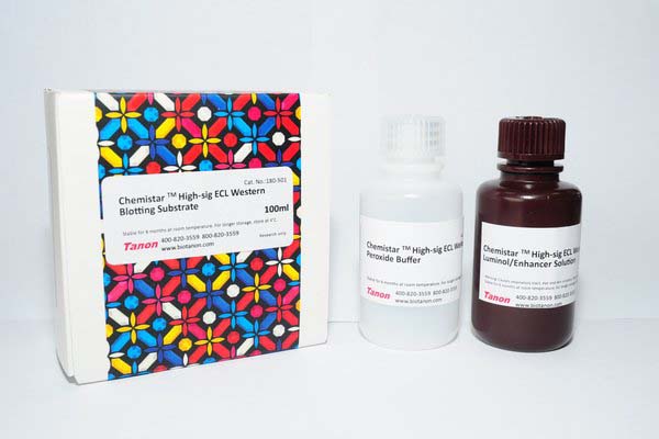
Chemistar™ High-sig ECL Western Blotting Substrate
|
Catalog No.
|
Size
|
|
180-501
|
100ml
|
|
180-5001
|
500ml
|
Chemistar™ High-sig ECL Western Blotting Substrate is a highly sensitive nonradioactive, enhanced luminol-based chemiluminescent substrate for the detection of horseradish peroxidase (HRP) on immunoblots. Tanon High-sig ECL Western Blotting Substrate enables the detection of picogram amounts of antigen and allows for easy detection of HRP using photographic or other imaging methods. Blots can be repeatedly exposed to X-ray film or Chemiluminescence imaging system to obtain optimal results or stripped of the immunodetection reagents and re-probed. The special formulation of Tanon High-sig ECL Western Blotting Substrate makes it the ideal substitute for Amersham ECL Substrate or Pierce ECL Western Blotting Substrate without the need for additional optimization of assay conditions.
Features
1. Use the same blotting conditions when switching from Amersham ECL Substrate or Pierce ECL Substrate to Tanon High-sig ECL Western Blotting Substrate.
2. If you are currently using a SuperSignal Chemiluminescent Substrate, switching to Tanon High-sig ECL Western Blotting Substrate requires increasing antigen and antibody concentrations. To determine the appropriate concentrations, perform a systematic dot blot analysis.
3. Tanon High-sig ECL Western Blotting Substrate requires more dilute antibody concentrations than those used with precipitating colorimetric HRP substrates. To optimize antibody concentrations, perform a systematic dot blot analysis.
4. Empirical testing is essential to determine the appropriate blocking reagent for each Western blot system, as cross-reactivity of the blocking reagent with antibodies causes nonspecific signal. Blocking buffer also affects system sensitivity.
5. Avoid using milk as a blocking reagent when using avidin/biotin systems because milk contains variable amounts of endogenous biotin, which causes high background signal.
6. Use sufficient volumes of wash buffer, blocking buffer, antibody solution and substrate working solution to cover blot and ensure that it never becomes dry. Using large blocking and wash buffer volumes minimizes nonspecific signal.
7. For optimal results, use a shaking platform during incubation steps.
8. Add Tween™-20 Detergent (final concentration of 0.05-0.1%) to the blocking buffer and all diluted antibody solutions to minimize nonspecific signal.
9. Do not use sodium azide as a preservative for buffers. Sodium azide is an inhibitor of HRP and could interfere with this system.
10. Do not handle membrane with bare hands. Always wear gloves or use clean forceps.
11. All equipment must be clean and free of foreign material. Metallic devices must have no visible signs of rust. Rust may cause speckling and high background.
12. Exposure to the sun or any other intense light can harm the substrate. For best results keep the substrate working solution in an amber bottle and avoid prolonged exposure to any intense light. Short-term exposure to typical laboratory lighting will not harm the working solution.
Store
1. Storage conditions: store at 2-8℃.
2. Dilute the primary antibody to 1:200-1:5000 from a 1mg/mL stock.
3. Dilute the secondary antibody to 1:2000-1:20000 from a 1mg/mL stock.
4. Mix Detection Reagents 1 and 2 at a 1:1 ratio and add it to the blot (Approximately 0.1mL of working HRP substrate is required per cm2 membrane area). Incubate blot for 1 minute.
5. Drain excess reagent. Cover blot with a clear plastic sheet protector or clear plastic wrap.
6. Expose blot to X -ray film or Chemiluminescence imaging system.
bio-equip.cn


